Lyme Disease infection may help trigger Lewy Body Dementia
Lyme Disease (LD) is a vector-borne disease that is transmitted to humans through exposure to the bacteria Borrelia burgdorferi (B. burgdorferi) from infected blacklegged ticks. Following a tick bite, symptoms like fatigue, headaches, skin rashes, and fever may develop. LD infection may also spread to vital organs such as the heart and nervous system, if left untreated1. Research has shown that B. burgdorferi can be found in post-mortem brain samples of patients who had been successfully treated for LD; evidence that the bacteria may persist in the central nervous system (CNS) and peripheral nervous system (PNS) tissue, even after antibiotic treatment2. Furthermore, it has been suggested that various infectious agents like Epstein Barr Virus, Herpes Simplex virus, C. pneumoniae, and B. burgdorferi may be associated with neurodegenerative disorders such as Parkinson’s disease, and that a rise in their infection burden correlates with increases in serum alpha synuclein (ASYN) levels3.
Previous Parkinson’s disease studies have shown that ASYN plays a pivotal role in modulating DNA folding and repair. One of the ways it does this is by interacting with nuclear histones and histone-free DNA. Investigating this phenomenon may be crucial in understanding the aggregation mechanisms of ASYN within Lewy bodies and neurites in various pathologies, including Diffuse Cortical Lewy Body Dementia (DCLBD).
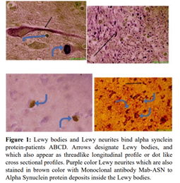
In a study written by Alan B. MacDonald in 2022, published in the Medical and Clinical Research Journal, it is hypothesized that since ASYN intrinsically binds human nuclear DNA, it may also complex with foreign DNA content found within Lewy bodies of DCLBD patients. This hypothesis was based on data observations acquired through DNA-focused research on a subset of four DCLBD patients. In the study, live B. burgdorferi bacteria, a causative agent of Lyme disease, was found to reside in Lewy brain neurons. Furthermore, MacDonald reported that B. burgdorferi proteins and extracellular DNA fragments were also specifically found inside Lewy neurites and Lewy bodies, respectively. To investigate this hypothesis, various techniques were employed. For example, StressMarq’s Alpha Synuclein (4F-1 clone) antibody (catalog#SMC-533) was used for immunohistochemistry to detect tissue-bound ASYN in Lewy bodies. Furthermore, Anti-OSP A (CB 10 clone) antibody, which was also used for immunohistochemistry, was combined with Fluorescence in situ Hybridization (FISH) and DNA intercalating stains. In conjunction with microscopy, those methods facilitated the detection of Borrelia-specific genes or deposits within Lewy bodies and demonstrated their co-localization with ASYN.
ASYN- bound Lewy bodies and neurites found in DCLBD patients
DCLBD pathogenesis was confirmed in patient samples with StressMarq’s Alpha Synuclein (4F-1 clone) antibody (catalog#SMC-533) by detecting the threshold amount of ASYN-positive Lewy body and Lewy neurites within a specific microscopic field of view. StressMarq’s Alpha Synuclein (3C11 clone) antibody (catalog# SMC-530) and Alpha Synuclein polyclonal antibody (catalog# SPC-800) were also used for validation purposes.

Taken from: Microbial DNA globular liquid crystal like deposits inside Lewy bodies in four Lewy dementia patients, MacDonald AB, Medical & Clinical Research. 2022;7(8):01-05
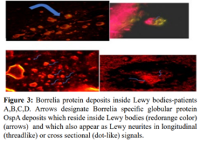
Borrelia infections deposit extracellular DNA and proteins inside Lewy neurons
Microscopic FISH hybridization data using FITC-conjugated probe bbO 0740, which is a validated sequence designed to anneal to microbial DNA in 30 strains of B.burgdorferi, shows that live Borrelia Spirochetes can be detected within the cytoplasm of cortical neurons adjacent to Lewy body structures. Additionally, immunostaining with Anti-OSP A (CB 10 clone) antibody confirms the presence of Borrelia-specific OSP A inside Lewy body and Lewy neurites within cortical neurons.
Cy5-conjugated probe bbO 0740 was also shown to anneal to Borrelia DNA deposits, originating from dead microbes, found within Lewy bodies. The microbial DNA deposits appear as globular liquid crystal formations. This was further confirmed using DNA intercalating staining with various dyes such as DAPI, Oligreen, Ethidium Bromide, and Acridine orange.

Taken from: Microbial DNA globular liquid crystal like deposits inside Lewy bodies in four Lewy dementia patients, MacDonald AB, Medical & Clinical Research. 2022;7(8):01-05
ASYN co-localizes with non-human DNA deposits within Lewy bodies and neurites
Live Borrelia bacteria were visualized inside Lewy neurons of DCLBD patients, and their extracellular DNA deposits were found within alpha synuclein-bound Lewy bodies and neurites. The co-localization of Borrelia DNA with alpha synuclein proteins is a newly explored phenomenon. Although alpha synuclein is a DNA-binding protein, its binding to foreign, non-human DNA may warrant future studies to explore the effect of DNA deposits, accrued by infectious agents within injured neurons on the aggregation mechanisms of alpha synuclein and formation of Lewy bodies and neurites in pathologies like Dementia and Parkinson’s disease.
Future perspectives
StressMarq is committed to supporting the research of Lewy Body Dementia (LBD) and Parkinson’s disease (PD) by providing researchers with constructs to establish in vitro and in vivo disease models, including alpha synuclein monomers, oligomers, pre-formed fibrils (PFFs), and alpha synuclein antibodies. Our fibrils are excellent for modeling disease pathology due to their ability to seed the aggregation of monomers. Meanwhile, the antibodies, as demonstrated by this study, are useful in helping detect tissue-bound alpha synuclein proteins within CNS tissue samples.
Article reference: Microbial DNA globular liquid crystal like deposits inside Lewy bodies in four Lewy dementia patients, MacDonald AB, Medical & Clinical Research. 2022;7(8):01-05.
References:
- Lyme Disease. (2022, January 19). Centers for Disease Control and Prevention (CDC). https://www.cdc.gov/lyme/index.html
- Detecting borrelia spirochetes: A case study with validation among autopsy specimens, Gadila, SKG et al., Frontiers in Neurology. 2021;12.
- The association between infectious burden and Parkinson’s disease: A case-control study, Bu XL et al., Parkinsonism & Related Disorders. 2015;21(8):877-881.


Leave a Reply