Featured Antibodies for Neurodegenerative Disease Research
StressMarq Biosciences has developed an extensive range of antibodies for use in neurodegenerative disease research to complement a cutting-edge portfolio of alpha synuclein, beta synuclein, gamma synuclein, tau, amyloid beta, SOD1 and TTR proteins. Our goal is to be the world leader in the development and supply of cutting-edge neuroscience research tools to assist scientists with disease model development and accelerate neurodegenerative disease drug discovery.
Alpha Synuclein Antibodies
Phosphorylated Alpha Synuclein
Ninety percent of alpha synuclein in Lewy bodies is phosphorylated at serine 129,1 making it a useful way to detect disease pathology in various synucleinopathies. Phosphorylation at serine 129 may also be involved in facilitating alpha synuclein aggregation and toxicity.1 StressMarq offers both monoclonal and polyclonal antibodies to pSer129 alpha synuclein.

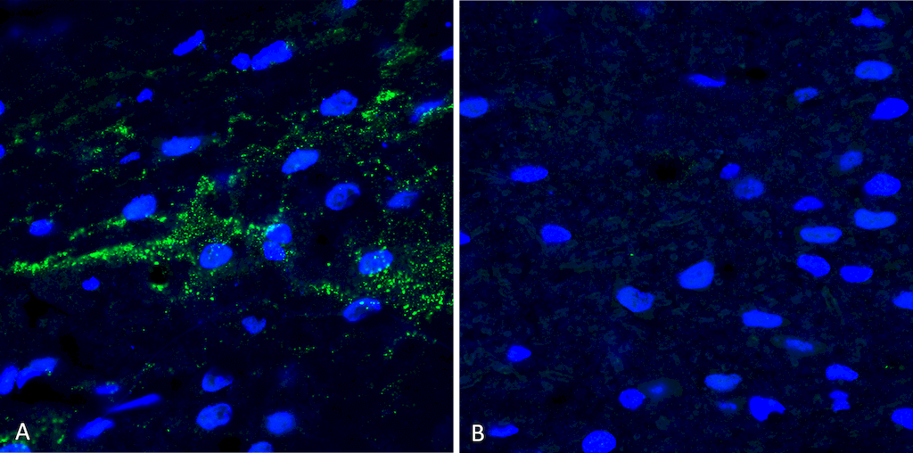
Immunohistochemistry analysis using StressMarq’s Anti-Alpha Synuclein (pSer129) Polyclonal Antibody (catalog# SPC-742). Mouse free floating brain sections. A) Right hemisphere (striatum) injected with alpha synuclein AAV vector. B) Control. Courtesy of: Trine Rasmussen, Simon Molgaard Jensen, Aarhus University.
![Rabbit Anti-Alpha Synuclein Antibody (pSer129) [J18] used in Immunocytochemistry/Immunofluorescence (ICC/IF) on Human iPSC-derived neurons (SMC-600)](https://www.stressmarq.com/wp-content/uploads/SMC-600_Alpha-Synuclein-pSer129_Antibody_J18_ICC-IF_Human_iPSC-derived-neurons_1-300x149.png)
Immunocytochemistry/Immunofluorescence analysis using StressMarq’s Anti-Alpha Synuclein (pSer129) Monoclonal Antibody, Clone J18 (catalog# SMC-600). Tissue: iPSC-derived neurons. Species: Human. Section A) negative control; no fibrils added to well. Section B) 7 days after addition of StressMarq’s Alpha Synuclein Pre-formed Fibrils (catalog# SPR-322).
Aggregated Alpha Synuclein
StressMarq’s Alpha Synuclein Aggregate Antibody [2F11] (catalog# SMC-617) binds a 3-dimensional epitope (including AA101-120 & AA61-870) present in MSA (Multiple System Atrophy) aggregates in human brain samples. ECL immunoblot analysis confirms detection and binding of alpha synuclein aggregates, and a lack of binding to monomeric alpha synuclein.
![Mouse Anti-Alpha Synuclein Antibody (Aggregate-Specific) [2F11] used in Immunohistochemistry (IHC) on Mouse gut (SMC-617)](https://www.stressmarq.com/wp-content/uploads/SMC-617_Alpha-Synuclein-Aggregate-Specific_Antibody_2F11_IHC_Mouse_gut_1-2-300x133.png)
Mouse gut samples stained with StressMarq’s Alpha Synuclein Aggregate Antibody [2F11] (catalog# SMC-617) at 1:1000 dilution with overnight incubation at 4°C in PBS with 0.1% BSA. Imaging at 40 x magnification. Mouse tissue used from a mouse Parkinson’s disease (PD) seeding model based on C57BLJ/6 mice. Top panel: Control. Bottom panel: Alpha Synuclein.
Phosphorylated Tau Antibodies
Tau hyperphosphorylation precedes tau aggregation in tauopathies such as Alzheimer’s disease, and can occur at a variety of phosphorylation sites.
Of particular interest is phosphorylation at Serine 202 and Threonine 205, which induces a turn-like structure and drives tau aggregation. StressMarq’s Tau (pSer202/pThr205) [AH36] Antibody (catalog# SMC-601) detects these phosphorylation sites, which are the same as those recognized by the AT8 tau antibody. Another phosphorylation site of interest is Threonine 217, detected by StressMarq’s Tau (pThr217) [15B7] (catalog# SMC-615).
![Rabbit Anti-Tau Antibody (pSer202/ pThr205) [AH36] used in Immunohistochemistry (IHC) on Mouse Brain slice (SMC-601)](https://www.stressmarq.com/wp-content/uploads/SMC-601_Tau_Antibody_AH36_IHC_Mouse_Brain-slice_1.png)
Immunohistochemistry analysis using StressMarq’s Tau (pSer202/pThr205) [AH36] Antibody (catalog# SMC-601) in mouse brain slices. Courtesy of: Julia Gerson
![Mouse Anti-Tau Antibody (pThr217) [15B7] used in Dot Blot (DB) on Human, Mouse, Rat Tau 2N4R P301S (phospho & non-phospho) (SMC-615)](https://www.stressmarq.com/wp-content/uploads/SMC-615_Tau_Antibody_15B7_DB_Human-Mouse-Rat_Tau-2N4R-P301S-phospho-non-phospho_1-2-300x205.png)
Dot Blot analysis of StressMarq’s Tau-441 (2N4R) P301S Mutant Pre-formed Fibrils expressed in E. coli (catalog# SPR-329) and Baculovirus/Sf9 (catalog# SPR-471). showing detection of Tau protein using Tau (pThr217) [15B7] Antibody (catalog# SMC-615). Color Development: Chemiluminescent for HRP (Moss) for 5 min in RT.
New! Amyloid Beta Oligomer Antibodies
The amyloid beta peptide (Aβ) is capable of aggregating into oligomers, protofibrils, and fibrils before forming insoluble plaques in the brain that facilitate disease pathology. Oligomeric amyloid beta has been observed to accumulate in the brain as early as one to two decades before the onset of Alzheimer’s disease symptoms. StressMarq is pleased to offer recombinant monoclonal antibodies targeting amyloid beta 1-42 oligomers as part of a diverse portfolio of research tools for neurodegenerative disease research.
![Rabbit Anti-Amyloid Beta 1-42 Oligomer Antibody [1A2] used in Dot Blot (DB) on Human Purified protein (SMC-618)](https://www.stressmarq.com/wp-content/uploads/SMC-618_Amyloid-Beta-1-42-Oligomer_Antibody_1A2_DB_Human_Purified-protein_1-300x207.png)
Dot blot analysis comparing detection of StressMarq’s Amyloid Beta, Alpha Synuclein and Tau protein constructs. Primary Antibodies: Amyloid Beta 1-42 Oligomer Antibody [1A2] (catalog# SMC-618), Alpha Synuclein Antibody (catalog# SPC-800), and Tau Antibody (catalog# SPC-801), all incubated for 1/2 hour at RT.
![Rabbit Anti-Amyloid Beta 1-42 Oligomer Antibody [2F9] used in Western Blot (WB) on Human Purified protein (SMC-620)](https://www.stressmarq.com/wp-content/uploads/SMC-620_Amyloid-Beta-1-42-Oligomer_Antibody_2F9_WB_Human_Purified-protein_1-300x232.png)
Western blot analysis comparing detection of StressMarq’s Amyloid Beta 1-42 constructs. Block: 5% skim milk. Primary Antibodies: Amyloid Beta 1-42 Oligomer Antibody [2F9] (catalog# SMC-620) & Amyloid Beta 1-16 Antibody [6E10] (Biolegend, 803001) at 1:10,000 & 1:1000 respectively, for 1/2 hour at RT.
References
- Sato, H., Kato, T. Arawaka, S. The role of Ser129 phosphorylation of α-synuclein in neurodegeneration of Parkinson’s disease: a review of in vivo models. Rev Neurosci. 2013.
