Discovery through Partnership | Excellence through Quality


Properties
| Storage Buffer | PBS pH7.2, 50% glycerol, 0.09% sodium azide *Storage buffer may change when conjugated |
| Storage Temperature | -20ºC, Conjugated antibodies should be stored according to the product label |
| Shipping Temperature | Blue Ice or 4ºC |
| Purification | Protein G Purified |
| Clonality | Monoclonal |
| Clone Number | 3A10.C9 |
| Isotype | IgG1 |
| Specificity | Detects ~20kDa (predicted mol.weight is ~21kDa). Does not cross-react with αA-crystallin, βL-crystallin, ΒH-crystallin, γ-crystallin, HSP25, HSP27 or HSP47 proteins. |
| Cite This Product | StressMarq Biosciences Cat# SMC-165, RRID: AB_2245270 |
| Certificate of Analysis | 0.5 µg/ml of SMC-165 was sufficient for detection of 50 ng purified alpha B crystalline by colorimetric immunoblot analysis using Goat anti-mouse IgG:HRP as the secondary antibody. |
Biological Description
| Alternative Names | AACRYA Antibody, Alpha crystallin B chain Antibody, CRYA2 Antibody, CRYAB Antibody, CTPP2 Antibody, HSPB5 Antibody, NY REN 27 antigen Antibody |
| Research Areas | Cancer, Cell Signaling, Chaperone Proteins, Heat Shock, Neuroscience, Protein Trafficking |
| Cellular Localization | Cytoplasm, Nucleus |
| Accession Number | NP_001876.1 |
| Gene ID | 1410 |
| Swiss Prot | P02511 |
| Scientific Background | The alpha-crystallins are major water-soluble lens structural proteins of the vertebrate eye that are related to the small heat shock protein family. The alpha-crystallins possess structural and functional similarities with HSP25 and HSP27 (1). Mammalian lens cystallins are divided into alpha, beta and gamma families. Alpha and beta families are further divided into acidic and basic groups (Alpha-A and Alpha-B respectively). In the lens, alpha-crystallin primarily functions to maintain proper refractive index, however it can also function as a molecular chaperone that binds to the denatured proteins, keeping them in solution and thereby maintaining the translucency of the lens. When cellular stress occurs, alpha-crystallin enters its' phosphorylated state and may serve a structural control function and play a role in protein maintenance (2). In addition to their interaction with proteins, alpha-crystallins also interact with native molecules such as membrane proteins, Golgi matrix protein, structural proteins, nuclear proteins and DNA (3, 4, 5, 6, and 7). Two other functions are an autokinase activity and participation in the intracellular architecture, and it has also been proven that both alpha-A and B prevent apoptosis by inhibiting caspases (8). Specifically, alpha-B cystallin is found in many cells and organs outside the lens, and alpha B is overexpressed in several neurological disorders and in cell lines under stress conditions (9). |
| References |
1. Merck K.B. et al. (1993) J Biol Chem. 268: 1046-1052. 2. Horwitz J. (1992) Proc Natl Acad Sci USA. 89(21): 10449-10453. 3. Cobb B.A. and Petrash J.M. (2002) Biochemistry. 41: 483-490 4. Horwitz J. (2003) Exp Eye Res. 76: 145-153. 5. Bullard B. et al. (2004) J Biol Chem. 279: 7917-7924. 6. Gangalum R.K., Schibler M.J. and Bhat S.P. (2004) J Biol Chem. 279: 43374.43377. 7. Maddala R. and Rao V.P. (2005) Exp Cell Res. 306: 203-215. 8. Yaung J., et al. (2007) Molecular Vision 13: 566-577. 9. Head M.W. et al. (2000) Neuropathol Appl Neurobiol. 26: 304-312. 10. Banduseela V.C., et al. (2009) Physiol Genomics. 39(3): 141-159. |
Product Images
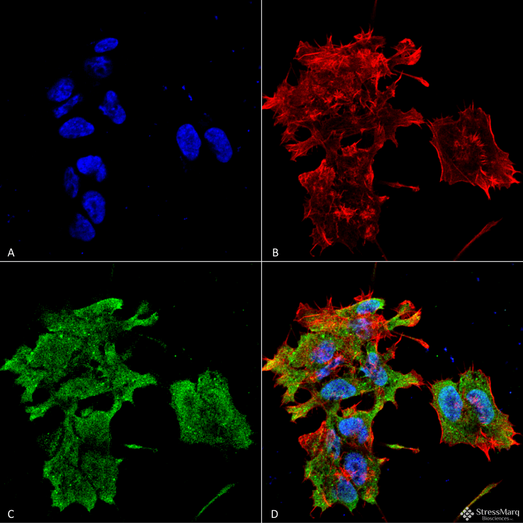
Immunocytochemistry/Immunofluorescence analysis using Mouse Anti-Alpha B Crystallin Monoclonal Antibody, Clone 3A10.C9 (SMC-165). Tissue: Neuroblastoma cell line (SK-N-BE). Species: Human. Fixation: 4% Formaldehyde for 15 min at RT. Primary Antibody: Mouse Anti-Alpha B Crystallin Monoclonal Antibody (SMC-165) at 1:100 for 60 min at RT. Secondary Antibody: Goat Anti-Mouse ATTO 488 at 1:100 for 60 min at RT. Counterstain: Phalloidin Texas Red F-Actin stain; DAPI (blue) nuclear stain at 1:1000, 1:5000 for 60min RT, 5min RT. Magnification: 60X. (A) DAPI (blue) nuclear stain. (B) Phalloidin Texas Red F-Actin stain. (C) Alpha B Crystallin Antibody. (D) Composite.
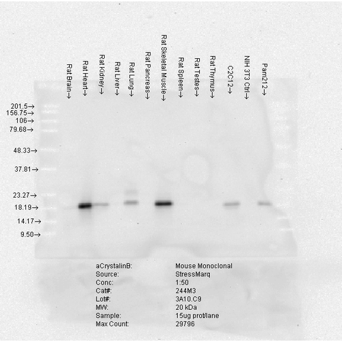
Western Blot analysis of Rat Brain, Heart, Kidney, Liver, Pancreas, Skeletal muscle, Spleen, Testes, Thymus cell lysates showing detection of Alpha B Crystallin protein using Mouse Anti-Alpha B Crystallin Monoclonal Antibody, Clone 3A10.C9 (SMC-165). Load: 15 µg. Block: 1.5% BSA for 30 minutes at RT. Primary Antibody: Mouse Anti-Alpha B Crystallin Monoclonal Antibody (SMC-165) at 1:50 for 2 hours at RT. Secondary Antibody: Sheep Anti-Mouse IgG: HRP for 1 hour at RT.
 Powered by Bioz
Evgeny Mymrikov :
Read the full review on pAbmAbs.com
StressMarq Biosciences :
Based on validation through cited publications.