Properties
| Storage Buffer | PBS pH 7.4, 50% glycerol, 0.09% Sodium azide *Storage buffer may change when conjugated |
| Storage Temperature | -20ºC, Conjugated antibodies should be stored according to the product label |
| Shipping Temperature | Blue Ice or 4ºC |
| Purification | Protein G Purified |
| Clonality | Monoclonal |
| Clone Number | 3C11 |
| Isotype | IgG1 |
| Specificity | Binds within the 81-130 aa region of human alpha synuclein, determined using WB and ELISA with corresponding peptides. Detects ~14 kDa band corresponding to Alpha synuclein. Band at ~30 kDa is a dimer. |
| Cite This Product | StressMarq Biosciences Cat# SMC-530, RRID: AB_2703053 |
| Certificate of Analysis | A 1:1000 dilution of SMC-530 was sufficient for detection of Alpha Synuclein in 15 µg of human brain cell lysate by ECL immunoblot analysis using goat anti-mouse IgG:HRP as the secondary antibody. |
Biological Description
| Alternative Names | Alpha Synuclein Antibody, Non-A beta component of AD amyloid Antibody, Non-A4 component of amyloid precursor Antibody, NACP Antibody, SNCA Antibody, PARK1 Antibody, PARK 1 Antibody, alphaSYN Antibody, PARK 4 antibody, PARK4 antibody, Parkinson disease familial 1 antibody, Parkinson disease (autosomal dominant, Lewy body) 4 antibody, SYN antibody |
| Research Areas | Alzheimer's Disease, Neurodegeneration, Neuroscience, Parkinson's Disease, Synuclein, Tangles & Tau, Multiple System Atrophy |
| Cellular Localization | Cell Junction, Cytoplasm, Cytosol, Membrane, Nucleus, Synapse |
| Accession Number | NP_000336.1 |
| Gene ID | 6622 |
| Swiss Prot | P37840 |
| Scientific Background | Alpha-Synuclein (SNCA) is expressed predominantly in the brain, where it is concentrated in presynaptic nerve terminals (1). Alpha-synuclein is highly expressed in the mitochondria of the olfactory bulb, hippocampus, striatum and thalamus (2). Functionally, it has been shown to significantly interact with tubulin (3), and may serve as a potential microtubule-associated protein. It has also been found to be essential for normal development of the cognitive functions; inactivation may lead to impaired spatial learning and working memory (4). SNCA fibrillar aggregates represent the major non A-beta component of Alzheimers disease amyloid plaque, and a major component of Lewy body inclusions, and Parkinson's disease. Parkinson's disease (PD) is a common neurodegenerative disorder characterized by the progressive accumulation in selected neurons of protein inclusions containing alpha-synuclein and ubiquitin (5, 6). |
| References |
1. “Genetics Home Reference: SNCA”. US National Library of Medicine. (2013). 2. Zhang L., et al. (2008) Brain Res. 1244: 40-52. 3. Alim M.A., et al. (2002) J Biol Chem. 277(3): 2112-2117. 4. Kokhan V.S., Afanasyeva M.A., Van'kin G. (2012) Behav. Brain. Res. 231(1): 226-230. 5. Spillantini M.G., et al. (1997) Nature. 388(6645): 839-840. 6. Mezey E., et al. (1998) Nat Med. 4(7): 755-757. |
Product Images
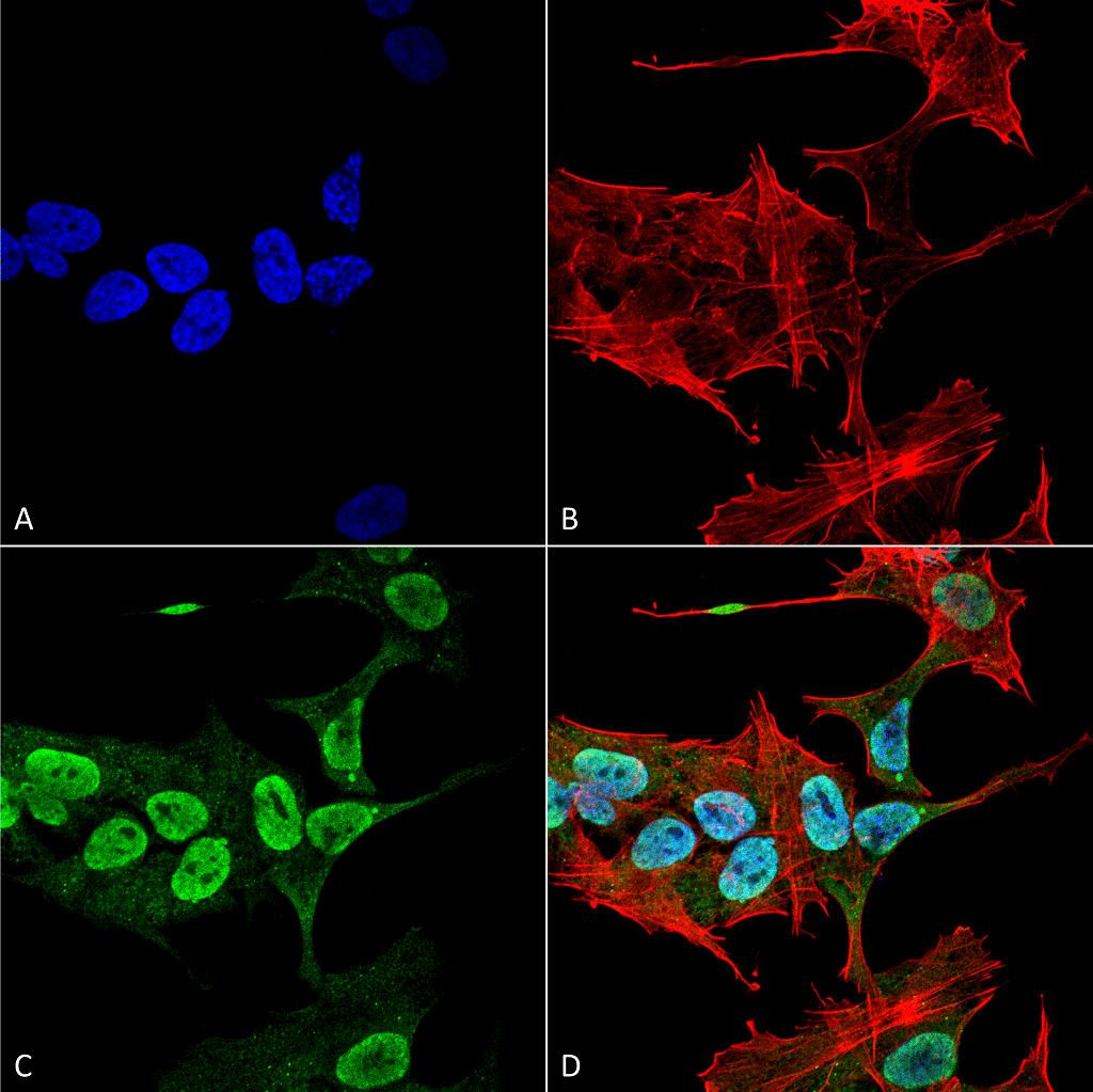
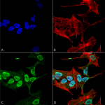
Immunocytochemistry/Immunofluorescence analysis using Mouse Anti-Alpha Synuclein Monoclonal Antibody, Clone 3C11 (SMC-530). Tissue: Neuroblastoma cell line (SK-N-BE). Species: Human. Fixation: 4% Formaldehyde for 15 min at RT. Primary Antibody: Mouse Anti-Alpha Synuclein Monoclonal Antibody (SMC-530) at 1:100 for 60 min at RT. Secondary Antibody: Goat Anti-Mouse ATTO 488 at 1:200 for 60 min at RT. Counterstain: Phalloidin Texas Red F-Actin stain; DAPI (blue) nuclear stain at 1:1000, 1:5000 for 60 min at RT, 5 min at RT. Localization: Cytoplasm: weak; Nucleus: Med. Magnification: 60X. (A) DAPI (blue) nuclear stain. (B) Phalloidin Texas Red F-Actin stain. (C) Alpha Synuclein Antibody. (D) Composite.
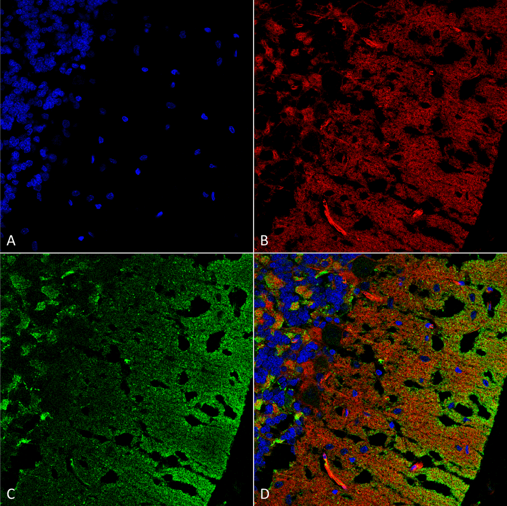
Immunohistochemistry analysis using Mouse Anti-Alpha Synuclein Monoclonal Antibody, Clone 3C11 (SMC-530). Tissue: cerebellum. Species: Rat. Fixation: Formalin fixed, paraffin embedded. Primary Antibody: Mouse Anti-Alpha Synuclein Monoclonal Antibody (SMC-530) at 1:25 for 1 hour at RT. Secondary Antibody: Goat Anti-Mouse IgG: Alexa Fluor 488. Counterstain: Actin-binding Phalloidin-Alexa Fluor 633; DAPI (blue) nuclear stain. Magnification: 63X. (A) DAPI (blue) nuclear stain. (B) Phalloidin Alexa Fluor 633 F-Actin stain. (C) Alpha Synuclein Antibody (D) Composite.
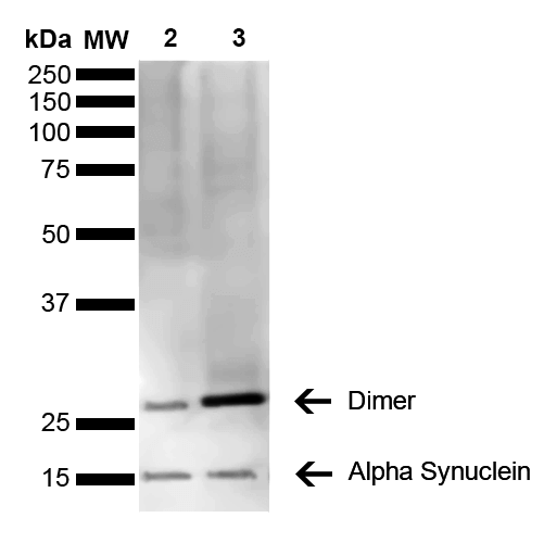
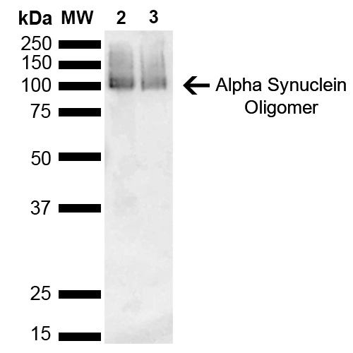
Western Blot analysis of Human Brain showing detection of 14 kDa Alpha Synuclein protein using Mouse Anti-Alpha Synuclein Monoclonal Antibody, Clone 3C11 (SMC-530). Lane 1: Molecular Weight Ladder (MW). Lane 2: Parkinson brain cell lystate. Lane 3: Human brain cell lysate. Load: 15 µg. Block: 5% Skim Milk in 1X TBST. Primary Antibody: Mouse Anti-Alpha Synuclein Monoclonal Antibody (SMC-530) at 1:1000 for 2 hours at RT. Secondary Antibody: Goat Anti-Mouse HRP:IgG at 1:3000 for 1 hour at RT. Color Development: ECL solution (Super Signal West Pico) for 5 min in RT. Predicted/Observed Size: 14 kDa. Other Band(s): 100 kDa (oligomer).
Western Blot analysis of Mouse, Rat Brain showing detection of 14 kDa Alpha Synuclein protein using Mouse Anti-Alpha Synuclein Monoclonal Antibody, Clone 3C11 (SMC-530). Lane 1: Molecular Weight Ladder (MW). Lane 2: Mouse brain cell lysate. Lane 3: Rat brain cell lysate. Load: 15 µg. Block: 5% Skim Milk in 1X TBST. Primary Antibody: Mouse Anti-Alpha Synuclein Monoclonal Antibody (SMC-530) at 1:1000 for 2 hours at RT. Secondary Antibody: Goat Anti-Mouse HRP:IgG at 1:3000 for 1 hour at RT. Color Development: ECL solution (Super Signal West Pico) for 5 min in RT. Predicted/Observed Size: 14 kDa. Other Band(s): ~30 kDa (dimer).
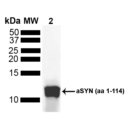
Western Blot analysis of Human Truncated Alpha Synuclein Protein showing detection of Alpha Synuclein protein using Mouse Anti-Alpha Synuclein Monoclonal Antibody, Clone 3C11 (SMC-530). Lane 1: MW Ladder. Lane 2: hASYN aa 1-114 (5 uL). Load: 5 uL. Block: 5% Skim Milk Powder in TBST. Primary Antibody: Mouse Anti-Alpha Synuclein Monoclonal Antibody (SMC-530) at 1:1000 for 2 hours at RT with shaking . Secondary Antibody: Goat anti-mouse IgG:HRP at 1:4000 for 1 hour at RT with shaking . Color Development: Chemiluminescent for HRP (Moss) for 5 min in RT.

![Mouse Anti-Alpha Synuclein Antibody [3C11] used in Immunohistochemistry (IHC) on Rat cerebellum (SMC-530)](https://www.stressmarq.com/wp-content/uploads/SMC-530_Alpha-Synuclein_Antibody_3C11_IHC_Rat_cerebellum_63X_1-100x100.png)
![Mouse Anti-Alpha Synuclein Antibody [3C11] used in Western Blot (WB) on Human Brain (SMC-530)](https://www.stressmarq.com/wp-content/uploads/SMC-530_Alpha-Synuclein_Antibody_3C11_WB_Human_Brain_1-100x100.png)
![Mouse Anti-Alpha Synuclein Antibody [3C11] used in Western Blot (WB) on Mouse, Rat Brain (SMC-530)](https://www.stressmarq.com/wp-content/uploads/SMC-530_Alpha-Synuclein_Antibody_3C11_WB_Mouse-Rat_Brain_1-100x100.png)
![Mouse Anti-Alpha Synuclein Antibody [3C11] used in Western Blot (WB) on Human Truncated Alpha Synuclein Protein (SMC-530)](https://www.stressmarq.com/wp-content/uploads/SMC-530_Alpha-Synuclein_Antibody_3C11_WB_Human_Truncated-Alpha-Synuclein-Protein_1-100x100.png)


StressMarq Biosciences :
Based on validation through cited publications.