Properties
| Storage Buffer | PBS pH7.4, 50% glycerol, 0.09% sodium azide *Storage buffer may change when conjugated |
| Storage Temperature | -20ºC, Conjugated antibodies should be stored according to the product label |
| Shipping Temperature | Blue Ice or 4ºC |
| Purification | Protein G Purified |
| Clonality | Monoclonal |
| Clone Number | N294A/6 (Formerly sold as S294A-6) |
| Isotype | IgG2b |
| Specificity | Detects ~140kDa (and smaller due to proteolytic cleavage). |
| Cite This Product | StressMarq Biosciences Cat# SMC-428, RRID: AB_2701451 |
| Certificate of Analysis | 1 µg/ml of SMC-428 was sufficient for detection of Brevican in 20 µg of rat brain lysate by colorimetric immunoblot analysis using Goat anti-mouse IgG:HRP as the secondary antibody. |
Biological Description
| Alternative Names | BCAN Antibody, BEHAB Antibody, CSPG7 Antibody, Brain enriched hyaluronan binding protein Antibody, Brevican core protein Antibody, Brevican core protein isoform 1 Antibody, Brevican core protein isoform 2 Antibody, Brevican proteoglycan Antibody, Chondroitin sulfate proteoglycan 7 Antibody, Chondroitin sulfate proteoglycan BEHAB Antibody, MGC13038 Antibody |
| Research Areas | Alzheimer's Disease, Cell Signaling, Neurodegeneration, Neuroscience, Scaffold Proteins |
| Cellular Localization | Extracellular Matrix, Extracellular Space, GPI-Anchor, Lipid-Anchor, Membrane |
| Accession Number | NP_001028837.1 |
| Gene ID | 25393 |
| Swiss Prot | P55068 |
| Scientific Background | Brevican is the most abundant chondroitin sulfate proteoglycan in the extracellular matrix of the adult brain. It is a member of the lectican family of aggregating extracellular matrix proteoglycans that bear chondroitin sulfate (CS) chains. It is highly expressed in the central nervous system and is thought to stabilize synapses and inhibit neural plasticity. Brevican is secreted from astrocytes and neurons as a 145 kD core protein that bears up to three, covalently-linked, CS chains. It is also is secreted as a 145 kD core protein without CS chains. When cleaved by extracellular glutamyl endopeptidases, the ADAMTSs, a 55 kD N-terminal fragment is formed that contains the unique C terminal epitope EAMESE. |
| References |
1. Yamada H., Watanabe K., Shimonaka M. and Yamaguchi Y. (1994) J. Biol. Chem. 269: 10119-10126. 2. Yamada H., Watanabe K., Shimonaka M., Yamasaki M. and Yamaguchi Y. (1995) Biochem. Biophys. Res. Commun. 216: 957-963. 3. Rauch U., et al. (1997) Genomics 44: 15-21. 4. Zhang H., Kelly G., Zerillo C., Jaworski D.M. and Hockfield S. (1998) J. Neurosci. 7: 2370-2376. 5. Aspberg A., Adam S., Kostka G., Timpl R. and Heinegard D. (1999) J. Biol. Chem. 274: 20444-20449. |
Product Images

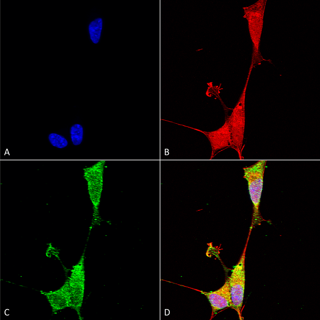
Immunocytochemistry/Immunofluorescence analysis using Mouse Anti-Brevican Monoclonal Antibody, Clone N294A/6 (SMC-428). Tissue: Neuroblastoma cells (SH-SY5Y). Species: Human. Fixation: 4% PFA for 15 min. Primary Antibody: Mouse Anti-Brevican Monoclonal Antibody (SMC-428) at 1:100 for overnight at 4°C with slow rocking. Secondary Antibody: AlexaFluor 488 at 1:1000 for 1 hour at RT. Counterstain: Phalloidin-iFluor 647 (red) F-Actin stain; Hoechst (blue) nuclear stain at 1:800, 1.6mM for 20 min at RT. (A) Hoechst (blue) nuclear stain. (B) Phalloidin-iFluor 647 (red) F-Actin stain. (C) Brevican Antibody (D) Composite.

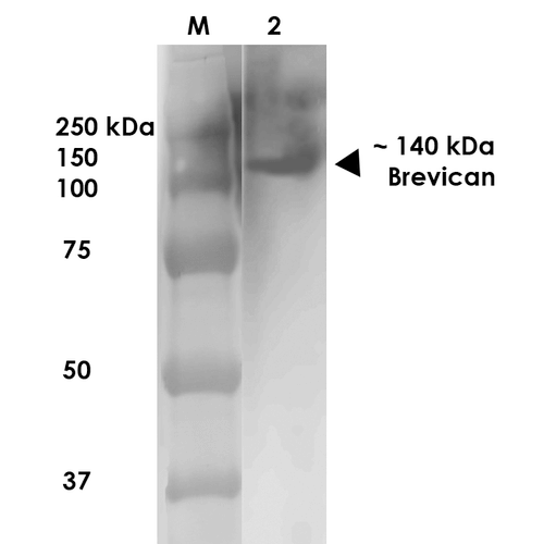
Western Blot analysis of Rat Brain Membrane showing detection of ~140 kDa Brevican protein using Mouse Anti-Brevican Monoclonal Antibody, Clone N294A/6 (SMC-428). Lane 1: MW Ladder. Lane 2: Rat Brain Membrane (10 µg). . Load: 10 µg. Block: 5% milk. Primary Antibody: Mouse Anti-Brevican Monoclonal Antibody (SMC-428) at 1:1000 for 1 hour at RT. Secondary Antibody: Goat Anti-Mouse IgG: HRP at 1:200 for 1 hour at RT. Color Development: TMB solution for 10 min at RT. Predicted/Observed Size: ~140 kDa.

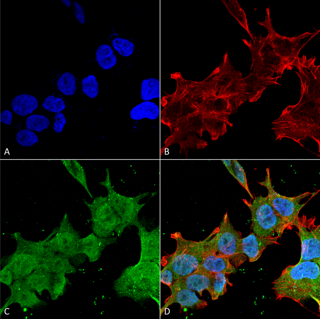
Immunocytochemistry/Immunofluorescence analysis using Mouse Anti-Brevican Monoclonal Antibody, Clone N294A/6 (SMC-428). Tissue: Neuroblastoma cell line (SK-N-BE). Species: Human. Fixation: 4% Formaldehyde for 15 min at RT. Primary Antibody: Mouse Anti-Brevican Monoclonal Antibody (SMC-428) at 1:100 for 60 min at RT. Secondary Antibody: Goat Anti-Mouse ATTO 488 at 1:100 for 60 min at RT. Counterstain: Phalloidin Texas Red F-Actin stain; DAPI (blue) nuclear stain at 1:1000, 1:5000 for 60min RT, 5min RT. Localization: Cell Membrane, Nucleus. Magnification: 60X. (A) DAPI (blue) nuclear stain. (B) Phalloidin Texas Red F-Actin stain. (C) Brevican Antibody. (D) Composite.


Reviews
There are no reviews yet.