Properties
| Storage Buffer | PBS pH7.4, 50% glycerol, 0.09% sodium azide *Storage buffer may change when conjugated |
| Storage Temperature | -20ºC, Conjugated antibodies should be stored according to the product label |
| Shipping Temperature | Blue Ice or 4ºC |
| Purification | Protein G Purified |
| Clonality | Monoclonal |
| Clone Number | N55/10 (Formerly sold as S55-10) |
| Isotype | IgG1 |
| Specificity | Detects ~260kDa. No cross-reactivity against Cav1.3. |
| Cite This Product | StressMarq Biosciences Cat# SMC-303, RRID: AB_2069540 |
| Certificate of Analysis | 1 µg/ml of SMC-303 was sufficient for detection of Cav3.2 in 10 µg of HEK cell lysate expressing Cav3.2 by colorimetric immunoblot analysis using Goat anti-mouse IgG:HRP as the secondary antibody. |
Biological Description
| Alternative Names | Cav3.2 Antibody, CACNA1H Antibody, CACNA1HB Antibody, calcium channel Antibody, voltage-dependent Antibody, T type Antibody, alpha 1H subunit Antibody, calcium channel Antibody, voltage-dependent Antibody, T type Antibody, alpha 1Hb subunit Antibody, ECA6 Antibody, EIG6 Antibody, FLJ90484 Antibody, Low-voltage-activated calcium channel alpha1 3.2 subunit Antibody, low-voltage-activated calcium channel alpha13.2 subunit Antibody, voltage dependent t-type calcium channel alpha-1H subunit Antibody, voltage-dependent T-type calcium channel subunit alpha-1H Antibody, voltage-gated calcium channel alpha subunit Cav3.2 Antibody, voltage-gated calcium channel alpha subunit CavT.2 Antibody, Voltage-gated calcium channel subunit alpha Cav3.2 Antibody |
| Research Areas | Calcium Channels, Cancer, Cell Signaling, Ion Channels, Neuroscience, Voltage-Gated Calcium Channels |
| Cellular Localization | Membrane |
| Accession Number | NP_001005407.1 |
| Gene ID | 8912 |
| Swiss Prot | O95180 |
| Scientific Background | CaV3.2 is a protein which in humans is encoded by the CACNA1H gene. Studies suggest certain mutations in this gene lead to childhood absence epilepsy (1, 2). Studies also suggest that the up-regulations of CaV3.2 may participate in the progression of prostate cancer toward an androgen-independent stage (3). |
| References |
1. Chen Y., et al. (2003) Ann. Neurol. 54(2): 239–43. 2. Khosravani H., et al. (2004) J Biol Chem. 279(11): 9681-9684. 3. Gackiere F., et al. (2008) J Biol Chem. 283(28): 19872. |
Product Images
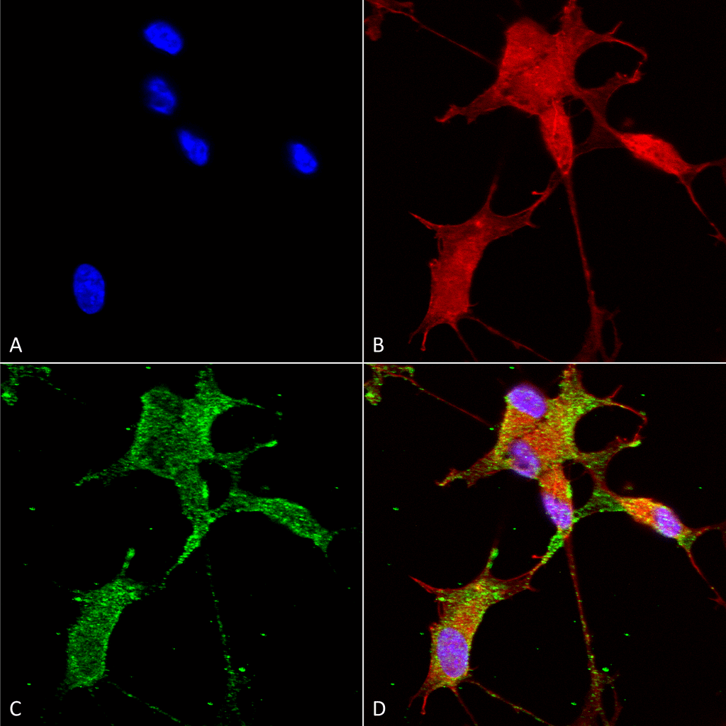
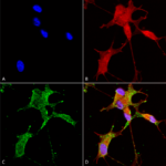
Immunocytochemistry/Immunofluorescence analysis using Mouse Anti-Cav3.2 Monoclonal Antibody, Clone N55/10 (SMC-303). Tissue: Neuroblastoma cells (SH-SY5Y). Species: Human. Fixation: 4% PFA for 15 min. Primary Antibody: Mouse Anti-Cav3.2 Monoclonal Antibody (SMC-303) at 1:50 for overnight at 4°C with slow rocking. Secondary Antibody: AlexaFluor 488 at 1:1000 for 1 hour at RT. Counterstain: Phalloidin-iFluor 647 (red) F-Actin stain; Hoechst (blue) nuclear stain at 1:800, 1.6mM for 20 min at RT. (A) Hoechst (blue) nuclear stain. (B) Phalloidin-iFluor 647 (red) F-Actin stain. (C) Cav3.2 Antibody (D) Composite.
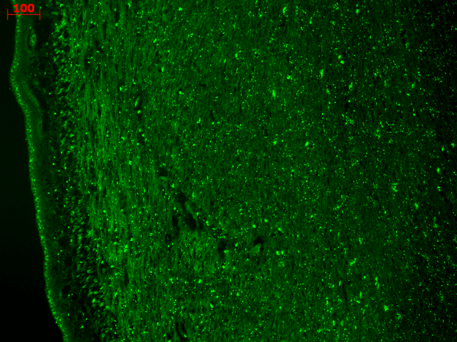
Immunohistochemistry analysis using Mouse Anti-CaV3.2 Calcium Channel Monoclonal Antibody, Clone N55/10 (SMC-303). Tissue: hippocampus. Species: Human. Fixation: Bouin’s Fixative and paraffin-embedded. Primary Antibody: Mouse Anti-CaV3.2 Calcium Channel Monoclonal Antibody (SMC-303) at 1:1000 for 1 hour at RT. Secondary Antibody: FITC Goat Anti-Mouse (green) at 1:50 for 1 hour at RT.
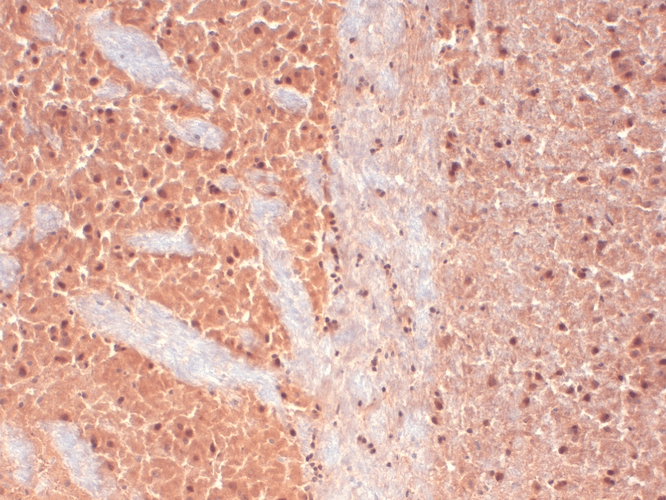
Immunohistochemistry analysis using Mouse Anti-CaV3.2 Calcium channel Monoclonal Antibody, Clone N55/10 (SMC-303). Tissue: frozen brain section. Species: Human. Fixation: 10% Formalin Solution for 12-24 hours at RT. Primary Antibody: Mouse Anti-CaV3.2 Calcium channel Monoclonal Antibody (SMC-303) at 1:1000 for 1 hour at RT. Secondary Antibody: HRP/DAB Detection System: Biotinylated Goat Anti-Mouse, Streptavidin Peroxidase, DAB Chromogen (brown) for 30 minutes at RT. Counterstain: Mayer Hematoxylin (purple/blue) nuclear stain at 250-500 µl for 5 minutes at RT.
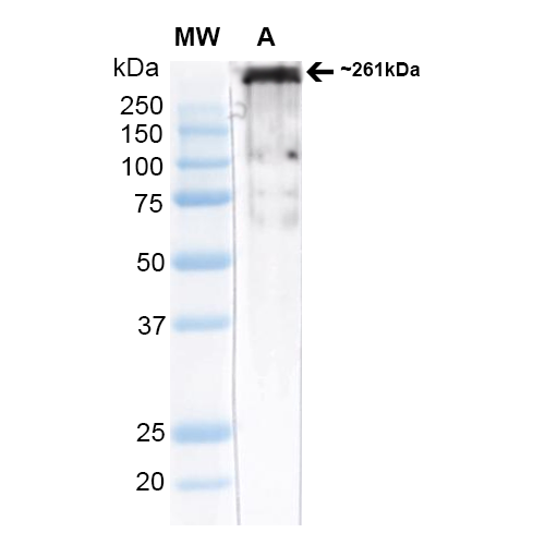
Western Blot analysis of Rat brain membrane lysate (native) showing detection of ~261 kDa Cav3.2 protein using Mouse Anti-Cav3.2 Monoclonal Antibody, Clone N55/10 (SMC-303). Block: 2% Skim Milk + 2% BSA in TBST. Primary Antibody: Mouse Anti-Cav3.2 Monoclonal Antibody (SMC-303) at 1:1000 for 2 hours at RT. Secondary Antibody: Anti-Mouse: HRP at 1:4000. Predicted/Observed Size: ~261 kDa.



StressMarq Biosciences :
Based on validation through cited publications.