Discovery through Partnership | Excellence through Quality


Properties
| Storage Buffer | PBS pH 7.4, 50% glycerol, 0.09% Sodium azide *Storage buffer may change when conjugated |
| Storage Temperature | -20ºC, Conjugated antibodies should be stored according to the product label |
| Shipping Temperature | Blue Ice or 4ºC |
| Purification | Protein G Purified |
| Clonality | Monoclonal |
| Clone Number | 10A6 |
| Isotype | IgG1 |
| Specificity | Specific for dityrosine modified proteins. Does not cross-react with 3,5-dibromotyrosine or bromotyrosine modified proteins. |
| Cite This Product | StressMarq Biosciences Cat# SMC-521, RRID: AB_2703017 |
| Certificate of Analysis | A 1:1000 dilution of SMC-521 was sufficient for detection of dityrosine in 1 µg of Dityrosine conjugated to BSA by ECL immunoblot analysis using Goat Anti-Mouse IgG:HRP as the secondary Antibody. |
Biological Description
| Alternative Names | Dityrosine Antibody, Dityrosine (DT) Antibody, DT Antibody, DY Antibody |
| Research Areas | Cancer, Cell Signaling, Oxidation, Oxidative Stress, Post-translational Modifications, Protein Oxidation |
| Scientific Background | ROS such as hydrogen peroxide (H2O2), superoxide, and hydroxyl radicals can react with both the backbone and the side chains of proteins, leading to backbone cleavage and side-chain modifications, respectively. Peroxidases, UV radiation, and hydroxyl radicals catalyze the formation of tyrosyl radicals which then react to form cross-links between proteins (1). This produces dityrosine, a metabolically stable biomarker of protein oxidation (2). |
| References |
1. Malencik, D. A., Anderson, S. R. (2003) Amino Acids. 25(3-4):233-247. 2. Giulivi, C., Davies, K. J. A. (2001) J Biol Chem. 276, 24129-24136. |
Product Images
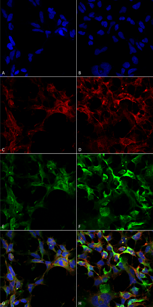
Immunocytochemistry/Immunofluorescence analysis using Mouse Anti-Dityrosine Monoclonal Antibody, Clone 10A6 (SMC-521). Tissue: Embryonic kidney epithelial cell line (HEK293). Species: Human. Fixation: 5% Formaldehyde for 5 min. Primary Antibody: Mouse Anti-Dityrosine Monoclonal Antibody (SMC-521) at 1:50 for 30-60 min at RT. Secondary Antibody: Goat Anti-Mouse Alexa Fluor 488 at 1:1500 for 30-60 min at RT. Counterstain: Phalloidin Alexa Fluor 633 F-Actin stain; DAPI (blue) nuclear stain at 1:250, 1:50000 for 30-60 min at RT. Localization: Cytoplasmic. Magnification: 20X (2X Zoom). (A,C,E,G) – Untreated. (B,D,F,H) – Cells cultured overnight with 50 µM H2O2. (A,B) DAPI (blue) nuclear stain. (C,D) Phalloidin Alexa Fluor 633 F-Actin stain. (E,F) Dityrosine Antibody. (G,H) Composite. Courtesy of: Dr. Robert Burke, University of Victoria.
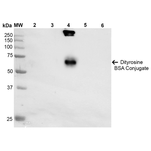
Western Blot analysis of Dityrosine-BSA Conjugate showing detection of 67 kDa Dityrosine protein using Mouse Anti-Dityrosine Monoclonal Antibody, Clone 10A6 (SMC-521). Lane 1: Molecular Weight Ladder (MW). Lane 2: BSA. Lane 3: 3,5-Dibromotyrosine-BSA. Lane 4: Dityrosine-BSA. Lane 5: Bromotyrosine-BSA. Lane 6: 7-ketocholesterol-BSA. Load: 1 µg. Block: 5% Skim Milk in TBST. Primary Antibody: Mouse Anti-Dityrosine Monoclonal Antibody (SMC-521) at 1:1000 for 2 hours at RT. Secondary Antibody: Goat Anti-Mouse IgG: HRP at 1:2000 for 60 min at RT. Color Development: ECL solution for 5 min in RT. Predicted/Observed Size: 67 kDa.
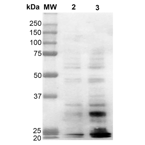
Western Blot analysis of Human Cervical cancer cell line (HeLa) lysate showing detection of Dityrosine protein using Mouse Anti-Dityrosine Monoclonal Antibody, Clone 10A6 (SMC-521). Lane 1: Molecular Weight Ladder (MW). Lane 2: HeLa cell lysate. Lane 3: H2O2 treated HeLa cell lysate. Load: 12 µg. Block: 5% Skim Milk in TBST. Primary Antibody: Mouse Anti-Dityrosine Monoclonal Antibody (SMC-521) at 1:1000 for 2 hours at RT. Secondary Antibody: Goat Anti-Mouse IgG: HRP at 1:2000 for 60 min at RT. Color Development: ECL solution for 5 min in RT.
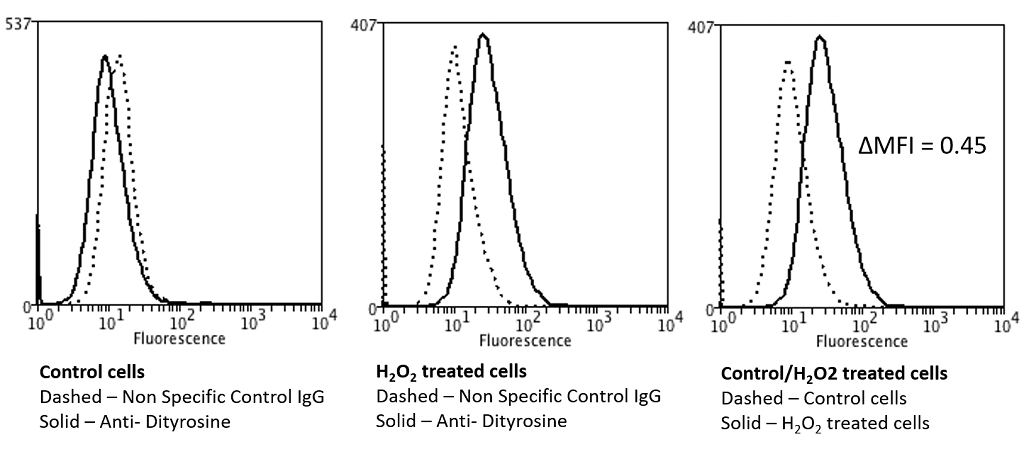
Flow Cytometry analysis using Mouse Anti-Dityrosine Monoclonal Antibody, Clone 10A6 (SMC-521). Tissue: Neuroblastoma cells (SH-SY5Y). Species: Human. Fixation: 90% Methanol. Primary Antibody: Mouse Anti-Dityrosine Monoclonal Antibody (SMC-521) at 1:50 for 30 min on ice. Secondary Antibody: Goat Anti-Mouse: PE at 1:100 for 20 min at RT. Isotype Control: Non Specific IgG. Cells were subject to oxidative stress by treating with 250 µM H2O2 for 24 hours.
 Powered by Bioz
Reviews
There are no reviews yet.