Properties
| Storage Buffer | PBS pH 7.4, 50% glycerol, 0.1% sodium azide *Storage buffer may change when conjugated |
| Storage Temperature | -20ºC, Conjugated antibodies should be stored according to the product label |
| Shipping Temperature | Blue Ice or 4ºC |
| Purification | Protein G Purified |
| Clonality | Monoclonal |
| Clone Number | N235/22 (Formerly sold as S235-22) |
| Isotype | IgG2b |
| Specificity | Detects ~30kDa. Does not cross-react with FGF13B/FHF2B. Cross reacts with FGF12A/FHF1A and FGF14A/FHF4A. |
| Cite This Product | StressMarq Biosciences Cat# SMC-448, RRID: AB_2701811 |
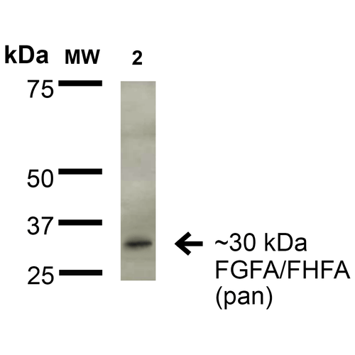
| Certificate of Analysis | 1 µg/ml of SMC-448 was sufficient for detection of FGFA/FHFA (pan) in 20 µg of rat brain lysate by colorimetric immunoblot analysis using Goat anti-mouse IgG:HRP as the secondary antibody. |
Biological Description
| Alternative Names | FGF-13 Antibody, FHF-2 Antibody, FGF13 Antibody, FHF2 Antibody, Acidic fibroblast growth factor Antibody, AFGF Antibody, Beta endothelial cell growth factor Antibody, Fibroblast growth factor homologous factor 2A Antibody, Fibroblast growth factor 13A Antibody, FGF13A Antibody, Beta-endothelial cell growth factor Antibody, ECGF Antibody, ECGFA Antibody, ECGFB Antibody, Endothelial Cell Growth Factor alpha Antibody, Endothelial Cell Growth Factor alpha Antibody, Endothelial Cell Growth Factor beta Antibody, FGF 1 Antibody, FGF alpha Antibody, FGFA Antibody, Fibroblast Growth Factor 1 Acidic Antibody, Fibroblast growth factor 1 Antibody, GLIO703 Antibody, HBGF 1 Antibody, HBGF1 Antibody, Heparin binding growth factor 1 Antibody, Heparin binding growth factor 1 precursor Antibody, Heparin-binding growth factor 1 Antibody |
| Research Areas | Angiogenesis, Cardiovascular Growth Factors, Cardiovascular System, FGF, Neurogenesis, Neuroscience |
| Cellular Localization | Cell projection, Cytoplasm, Dendrite, Growth cone, Nucleus |
| Accession Number | AAH34340 |
| Gene ID | 2258 |
| Swiss Prot | Q92913 |
| Scientific Background | FGF13(Fibroblast growth factor 13), also called FHF2 is a protein that in humans is encoded by the FGF13 gene.The protein encoded by this gene is a member of the fibroblast growth factor (FGF) family. FGF13is a large gene, extending over approximately 200 kb in Xq26.3, and contains at least 7 exons. By cytogenetic, FISH, and database analysis, Gecz et al. (1999) localized the FGF13 gene within a 400-kb duplication interval on chromosome Xq26.3. FGF family members possess broad mitogenic and cell survival activities, and are involved in a variety of biological processes, including embryonic development, cell growth, morphogenesis, tissue repair, tumor growth, and invasion. This gene is located to a region associated with Borjeson-Forssman-Lehmann syndrome (BFLS), a syndromal X-linked mental retardation, which suggests it may be a candidate gene for familial cases of the BFL syndrome. The function of this gene has not yet been determined. Two alternatively spliced transcripts encoding different isoforms have been described for this gene. |
| References |
1. Gecz, J., et al. (1999) Hum. Genet. 104: 56-63. 2. Smallwood, P. M., et al. (1996) Proc. Nat. Acad. Sci. 93: 9850-9857. |
Product Images

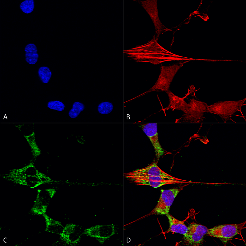
Immunocytochemistry/Immunofluorescence analysis using Mouse Anti-FGFA/FHFA (pan) Monoclonal Antibody, Clone N235/22 (SMC-448). Tissue: Neuroblastoma cells (SH-SY5Y). Species: Human. Fixation: 4% PFA for 15 min. Primary Antibody: Mouse Anti-FGFA/FHFA (pan) Monoclonal Antibody (SMC-448) at 1:50 for overnight at 4°C with slow rocking. Secondary Antibody: AlexaFluor 488 at 1:1000 for 1 hour at RT. Counterstain: Phalloidin-iFluor 647 (red) F-Actin stain; Hoechst (blue) nuclear stain at 1:800, 1.6mM for 20 min at RT. (A) Hoechst (blue) nuclear stain. (B) Phalloidin-iFluor 647 (red) F-Actin stain. (C) FGFA/FHFA (pan) Antibody (D) Composite.

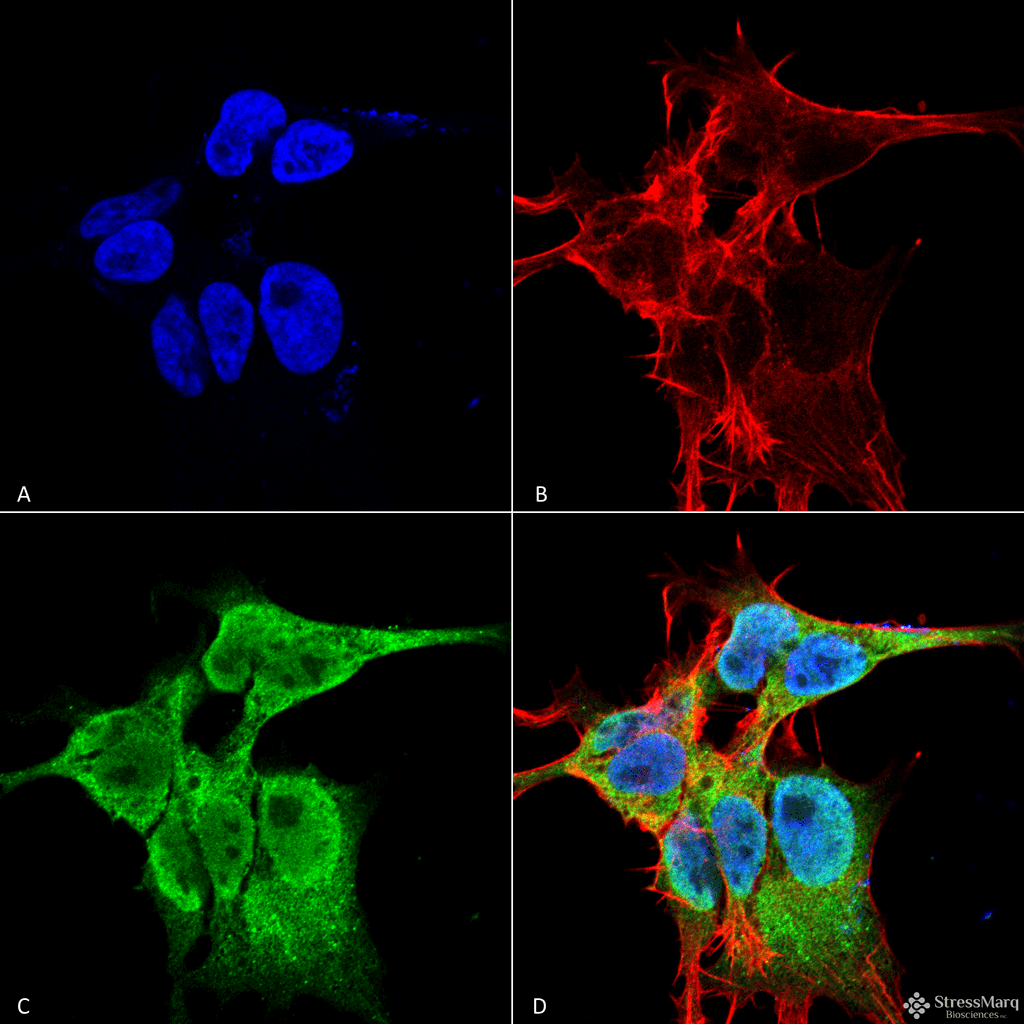
Immunocytochemistry/Immunofluorescence analysis using Mouse Anti-FGFA/FHFA (pan) Monoclonal Antibody, Clone N235/22 (SMC-448). Tissue: Neuroblastoma cell line (SK-N-BE). Species: Human. Fixation: 4% Formaldehyde for 15 min at RT. Primary Antibody: Mouse Anti-FGFA/FHFA (pan) Monoclonal Antibody (SMC-448) at 1:100 for 60 min at RT. Secondary Antibody: Goat Anti-Mouse ATTO 488 at 1:100 for 60 min at RT. Counterstain: Phalloidin Texas Red F-Actin stain; DAPI (blue) nuclear stain at 1:1000; 1:5000 for 60 min RT, 5 min RT. Localization: Cell Projection, Nucleus, Cytoplasm. Magnification: 60X. (A) DAPI (blue) nuclear stain. (B) Phalloidin Texas Red F-Actin stain. (C) FGFA/FHFA (pan) Antibody. (D) Composite.

Western Blot analysis of Rat Brain Membrane showing detection of ~30 kDa FGFA/FHFA (pan) protein using Mouse Anti-FGFA/FHFA (pan) Monoclonal Antibody, Clone N235/22 (SMC-448). Lane 1: Molecular Weight Ladder. Lane 2: Rat Brain Membrane. Load: 15 µg. Block: 2% BSA and 2% Skim Milk in 1X TBST. Primary Antibody: Mouse Anti-FGFA/FHFA (pan) Monoclonal Antibody (SMC-448) at 1:200 for 16 hours at 4°C. Secondary Antibody: Goat Anti-Mouse IgG: HRP at 1:1000 for 1 hour RT. Color Development: ECL solution for 6 min in RT. Predicted/Observed Size: ~30 kDa.


Reviews
There are no reviews yet.