Properties
| Storage Buffer | PBS pH 7.4, 50% glycerol, 0.1% sodium azide *Storage buffer may change when conjugated |
| Storage Temperature | -20ºC, Conjugated antibodies should be stored according to the product label |
| Shipping Temperature | Blue Ice or 4ºC |
| Purification | Protein G Purified |
| Clonality | Monoclonal |
| Clone Number | N355/1 (Formerly sold as S355-1) |
| Isotype | IgG1 |
| Specificity | Detects ~100kDa. Does not cross-react with GluR2. |
| Cite This Product | StressMarq Biosciences Cat# SMC-440, RRID: AB_2701667 |
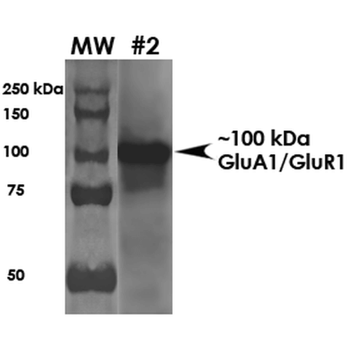
| Certificate of Analysis | 1 µg/ml of SMC-440 was sufficient for detection of GluA1/GluR1 in 20 µg of mouse brain membrane lysate and assayed by colorimetric immunoblot analysis using goat anti-mouse IgG:HRP as the secondary antibody. |
Biological Description
| Alternative Names | AMPA 1 Antibody, AMPA selective glutamate receptor 1 Antibody, Glutamate receptor 1 Antibody, GluA1 Antibody, GLUH 1 Antibody, GluR K1 Antibody, GluRK1 Antibody, Glr-1 Antibody, Glur-1 Antibody, HIPA Antibody, Glur1 Antibody, GluR-A Antibody, AI853806 Antibody, 2900051M01Rik Antibody, GluRA Antibody, Glr1 Antibody, MGC13325 Antibody, GLUH1 Antibody, GluA1 Antibody, GLUR1 Antibody, HBGR1 Antibody, GLURA Antibody, gluR- Antibody, GluA1 Antibody, Glutamate receptor ionotropic AMPA 1 Antibody, Glutamate receptor ionotropic Antibody, Gria 1 Antibody, HBGR1 Antibody, OTTHUMP00000160643 Antibody, OTTHUMP00000165781 Antibody, OTTHUMP00000224241 Antibody, OTTHUMP00000224242 Antibody, OTTHUMP00000224243 Antibody |
| Research Areas | AMPA Receptors, Cell Signaling, Glutamate Receptors, Neuroscience, Neurotransmitter Receptors |
| Cellular Localization | Cell Junction, Cell membrane, Endoplasmic Reticulum |
| Accession Number | NP_113796 |
| Gene ID | 50592 |
| Swiss Prot | P19490 |
| Scientific Background | Glutamic acid is the major excitatory neurotransmitter in the mammalian central nervous system. Glutamate receptors are classified on the basis of their activation by different agonists (1-3). GluR1, human glutamate receptor type 1, is an integral membrane protein that is widely expressed in the human brain. The postsynaptic actions of glutamic acid are mediated by a variety of receptors that are named according to their selective agonists. GluR1 is known to bind a kainate subtype of agonist. It has been found that malfunctioning of the glutamatergic system may result in certain brain disorders and neurodegeneration (3). |
| References |
1. Potier M.C., Spillantini M.G., Carter N.P. (1992) DNA Seq. 2(4): 211-218. 2. Puckett, C., et al. (1991) Proc. Nat. Acad. Sci. USA 88(17): 7557-7561. 3. Gregor, P., et al. (1993) Proc. Nat. Acad. Sci. USA 90: 3053-3057. |
Product Images

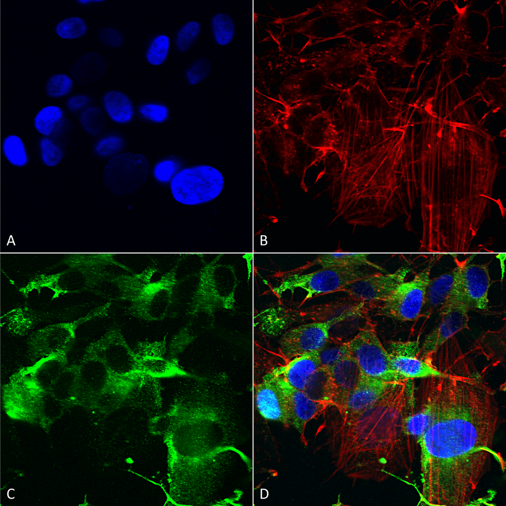
Immunocytochemistry/Immunofluorescence analysis using Mouse Anti-GluA1/GluR1 Monoclonal Antibody, Clone N355/1 (SMC-440). Tissue: Neuroblastoma cells (SH-SY5Y). Species: Human. Fixation: 4% PFA for 15 min. Primary Antibody: Mouse Anti-GluA1/GluR1 Monoclonal Antibody (SMC-440) at 1:200 for overnight at 4°C with slow rocking. Secondary Antibody: AlexaFluor 488 at 1:1000 for 1 hour at RT. Counterstain: Phalloidin-iFluor 647 (red) F-Actin stain; Hoechst (blue) nuclear stain at 1:800, 1.6mM for 20 min at RT. (A) Hoechst (blue) nuclear stain. (B) Phalloidin-iFluor 647 (red) F-Actin stain. (C) GluA1/GluR1 Antibody (D) Composite.

Western Blot analysis of Rat Brain Membrane showing detection of ~100 kDa GluA1-GluR1 protein using Mouse Anti-GluA1-GluR1 Monoclonal Antibody, Clone N355/1 (SMC-440). Load: 10 µg. Block: 5% milk + TBST. Primary Antibody: Mouse Anti-GluA1-GluR1 Monoclonal Antibody (SMC-440) at 1:2000 for 1 hour at RT. Secondary Antibody: Goat Anti-Mouse HRP at 1:200 for 1 hour at RT. Predicted/Observed Size: ~100 kDa.

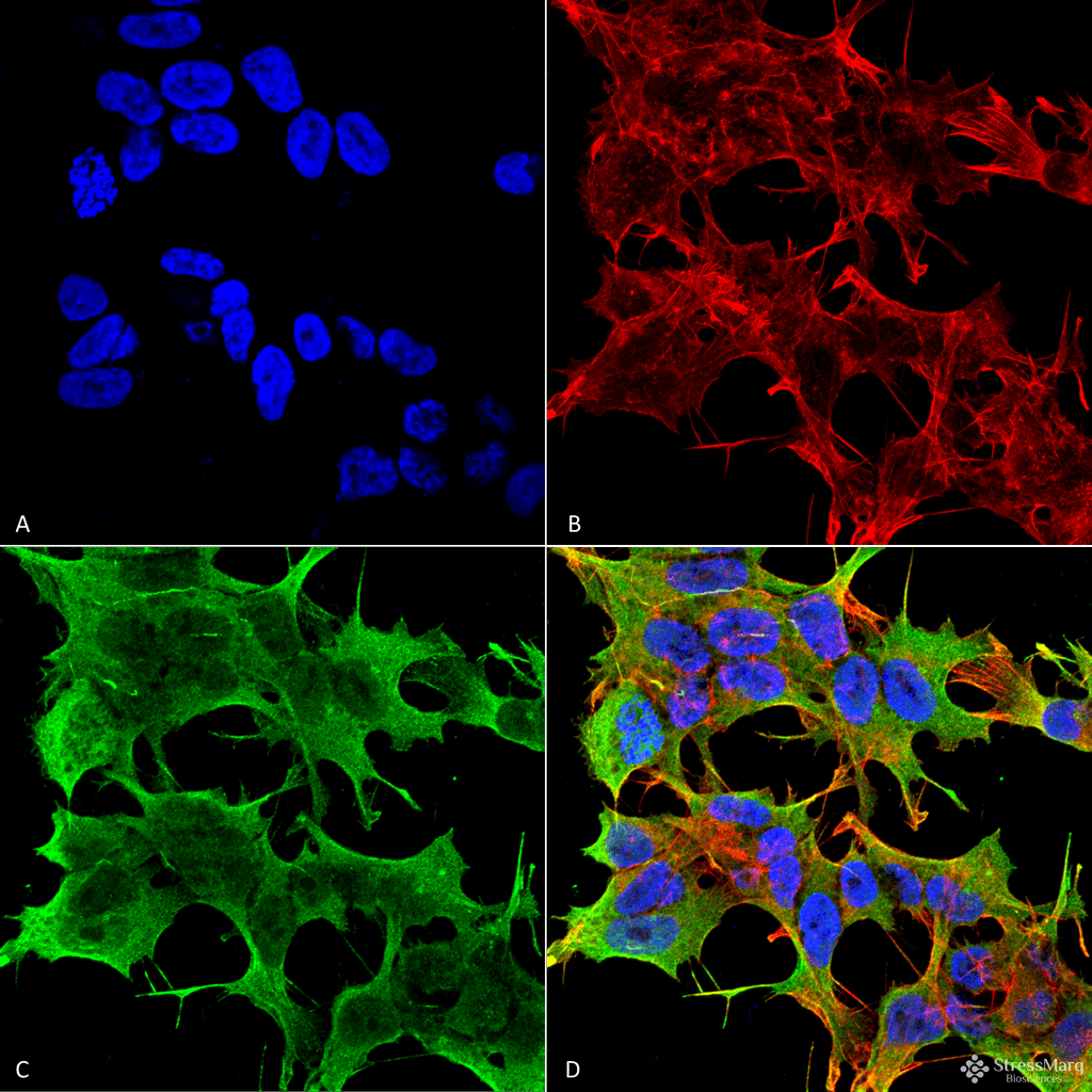
Immunocytochemistry/Immunofluorescence analysis using Mouse Anti-GluA1/GluR1 Glutamate Receptor Monoclonal Antibody, Clone N355/1 (SMC-440). Tissue: Neuroblastoma cell line (SK-N-BE). Species: Human. Fixation: 4% Formaldehyde for 15 min at RT. Primary Antibody: Mouse Anti-GluA1/GluR1 Glutamate Receptor Monoclonal Antibody (SMC-440) at 1:100 for 60 min at RT. Secondary Antibody: Goat Anti-Mouse ATTO 488 at 1:100 for 60 min at RT. Counterstain: Phalloidin Texas Red F-Actin stain; DAPI (blue) nuclear stain at 1:1000, 1:5000 for 60min RT, 5min RT. Localization: Cell Membrane, Cell Junction. Magnification: 60X. (A) DAPI (blue) nuclear stain. (B) Phalloidin Texas Red F-Actin stain. (C) GluA1/GluR1 Glutamate Receptor Antibody. (D) Composite.


Reviews
There are no reviews yet.