Discovery through Partnership | Excellence through Quality


Properties
| Storage Buffer | PBS pH 7.4, 50% glycerol, 0.1% sodium azide *Storage buffer may change when conjugated |
| Storage Temperature | -20ºC, Conjugated antibodies should be stored according to the product label |
| Shipping Temperature | Blue Ice or 4ºC |
| Purification | Protein G Purified |
| Clonality | Monoclonal |
| Clone Number | 4B4 |
| Isotype | IgG1 |
| Specificity | Detects ~85kDa (unstressed cell lysates) and ~95kDa (heat shocked cell lysates). |
| Cite This Product | StressMarq Biosciences Cat# SMC-477, RRID: AB_2702297 |
| Certificate of Analysis | 1 µg/ml of SMC-477 was sufficient for detection of HSF1 in 20 µg of heat shocked HeLa cell lysate by colorimetric immunoblot analysis using Rabbit anti-rat IgG: AP as the secondary antibody. |
Biological Description
| Alternative Names | HSTF1 Antibody, Heat shock factor protein 1 Antibody, Heat shock transcription factor 1 Antibody, HSF 1 Antibody |
| Research Areas | Cancer, Cardiovascular System, Cell Signaling, Epigenetics and Nuclear Signaling, Heart, Heat Shock |
| Cellular Localization | Cytoplasm, Nucleus |
| Accession Number | NP_032322.1 |
| Gene ID | 15499 |
| Swiss Prot | P38532 |
| Scientific Background | HSF1, or heat shock factor 1, belongs to a family of Heat Shock transcription factors that activate the transcription of genes encoding products required for protein folding, processing, targeting, degradation, and function (2). The up-regulation of HSP (heat shock proteins) expression by stressors is achieved at the level of transcription through a heat shock element (HSE) and a transcription factor (HSF) (3, 4, 5). Most HSFs have highly conserved amino acid sequences. On all HSFs there is a DNA binding domain at the N-terminus. Hydrophobic repeats located adjacent to this binding domain are essential for the formation of active trimers. Towards the C-terminal region another short hydrophobic repeat exists, and is thought to be necessary for suppression of trimerization (6). There are two main heat shock factors, 1 and 2. Mouse HSF1 exists as two isoforms, however in higher eukaryotes HSF1 is found in a diffuse cytoplasmic and nuclear distribution in un-stressed cells. Once exposed to a multitude of stressors, it localizes to discrete nuclear granules within seconds. As it recovers from stress, HSF1 dissipates from these granules to a diffuse nuceloplasmic distribution. HSF2 on the other hand is similar to mouse HSF1, as it exists as two isoforms, the alpha form being more transcriptionally active than the smaller beta form (7, 8). Various experiments have suggested that HFS2 may have roles in differentiation and development (9, 10, 11). |
| References |
1. Cotto J.J., Fox S.G. and Morimoto R.I. (1997) J. Cell Science 110: 2925-2934. 2. Morano K.A. and Thiele D.J. (1999). Gene Expression 7 (6): 271-82. 3. Tanaka K.I., et al. (2007). JBC Papers Online Manuscript M704081200. 4. Morimoto R. I. (1998) Genes Dev 12: 3788-3796. 5. McMillan D. R., et al. (1998) J Bio Chem 273: 7523-7528. 6. Jolly C., Usson Y. and Morimoto R.I. (1999) Proc. Natl. Acad. Sci. USA 96 (12): 6769- 6774. 7. Fiorenza M.T., et al. (1995) Nucleic Acids Res. 23 (3):467-474. 8. Goodson M.L., Park-Sarge O.K. and Sarge K.D. (1995) Mol. Cell. Biol. 15(10): 5288-5293. 9. Rallu M., et al. (1997) Proc. Natl. Acad. Sci. USA 94(6): 2392-2397. 10. Sarge K.D., et al. (1994) Biol. Reprod. 50(6): 1334-1343. 11. Murphy S.P., Gorzowski J.J., Sarge K.D. and Phillips B. (1994) Mol. Cell. Biol. 14(8):5309-5317. |
Product Images
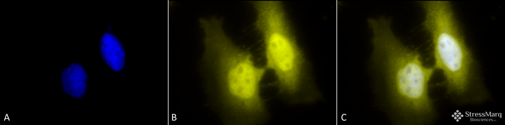
Immunocytochemistry/Immunofluorescence analysis using Rat Anti-HSF1 Monoclonal Antibody, Clone 4B4 (SMC-477). Tissue: Heat Shocked cervical cancer cells (HeLa). Species: Human. Fixation: 2% Formaldehyde for 20 min at RT. Primary Antibody: Rat Anti-HSF1 Monoclonal Antibody (SMC-477) at 1:100 for 12 hours at 4°C. Secondary Antibody: R-PE Goat Anti-Rat (yellow) at 1:200 for 2 hours at RT. Counterstain: DAPI (blue) nuclear stain at 1:40000 for 2 hours at RT. Localization: Cytoplasm. Localizes to the nucleus upon activation. Magnification: 100x. (A) DAPI (blue) nuclear stain. (B) Anti-HSF1 Antibody. (C) Composite. Heat Shocked at 42°C for 1h.
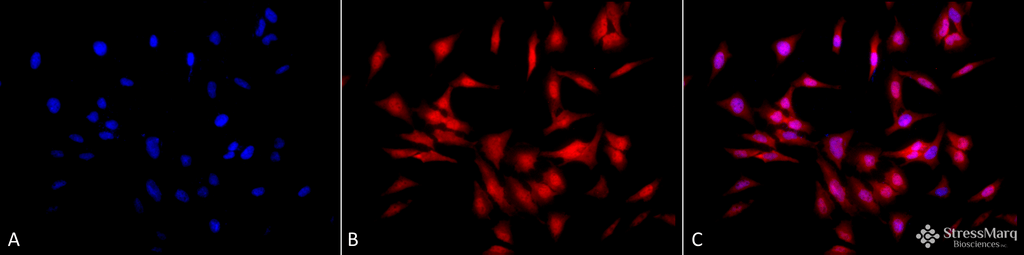
Immunocytochemistry/Immunofluorescence analysis using Rat Anti-HSF1 Monoclonal Antibody, Clone 4B4 (SMC-477). Tissue: Heat Shocked cervical cancer cells (HeLa). Species: Human. Fixation: 2% Formaldehyde for 20 min at RT. Primary Antibody: Rat Anti-HSF1 Monoclonal Antibody (SMC-477) at 1:100 for 12 hours at 4°C. Secondary Antibody: APC Goat Anti-Rat (red) at 1:200 for 2 hours at RT. Counterstain: DAPI (blue) nuclear stain at 1:40000 for 2 hours at RT. Localization: Cytoplasm. Localizes to the nucleus upon activation. Magnification: 20x. (A) DAPI (blue) nuclear stain. (B) Anti-HSF1 Antibody. (C) Composite. Heat Shocked at 42°C for 1h.
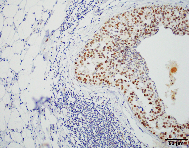
Immunohistochemistry analysis using Rat Anti-HSF1 Monoclonal Antibody, Clone 4B4 (SMC-477). Tissue: Breast carcinoma. Species: Human. Fixation: 10% Formalin Solution for 20 hours at RT. Primary Antibody: Rat Anti-HSF1 Monoclonal Antibody (SMC-477) at 1:2000 for 40 min. Secondary Antibody: Dako labeled Polymer HRP Anti-rat IgG, DAB Chromogen (brown) (Dako Envision+ System) for 30 min at RT. Counterstain: Mayer’s Hematoxylin (purple/blue) nuclear stain for 1 minute at RT. Localization: Nuclear. Magnification: 100X. Courtesy of: Dr. Sandro Santagata, Harvard Medical School.
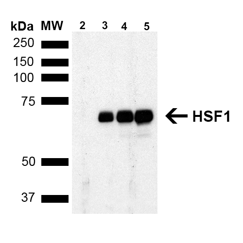
Western Blot analysis of Human Breast adenocarcinoma cell line (MCF7) showing detection of ~65 kDa HSF1 protein using Rat Anti-HSF1 Monoclonal Antibody, Clone 4B4 (SMC-477). Lane 1: MW ladder. Lane 2: HSF1 null lysate prepared from mouse embryonic fibroblasts. Lane 3: MCF7 lysate (5 µg). Lane 4: MCF7 lysate (10 µg). Lane 5: MCF7 lysate (20 µg). Block: 1.5% BSA for 30 minutes at RT. Primary Antibody: Rat Anti-HSF1 Monoclonal Antibody (SMC-477) at 1:1000 for 2 hours at RT. Secondary Antibody: Goat Anti-Rat IgG: HRP for 1 hour at RT. Predicted/Observed Size: ~65 kDa. Courtesy of: Dr. Sandro Santagata, Harvard Medical School.
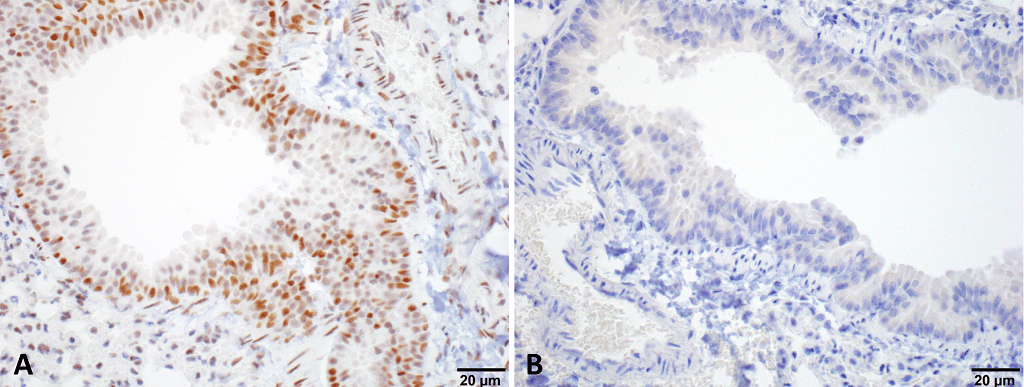
Immunohistochemistry analysis using Rat Anti-HSF1 Monoclonal Antibody, Clone 4B4 (SMC-477). Tissue: Lung. Species: Mouse. Fixation: 10% Formalin Solution for 20 hours at RT. Primary Antibody: Rat Anti-HSF1 Monoclonal Antibody (SMC-477) at 1:1000 for 40 min. Secondary Antibody: Dako labeled Polymer HRP Anti-rat IgG, DAB Chromogen (brown) (Dako Envision+ System) for 30 min at RT. Counterstain: Mayer’s Hematoxylin (purple/blue) nuclear stain for 1 minute at RT. Localization: Nuclear. Magnification: 100X. (A) HSF Wildtype. (B) HSF null. Courtesy of: Dr. Sandro Santagata, Harvard Medical School.
 Powered by Bioz
Reviews
There are no reviews yet.