Discovery through Partnership | Excellence through Quality


Properties
| Storage Buffer | PBS pH7.2, 50% glycerol, 0.09% sodium azide *Storage buffer may change when conjugated |
| Storage Temperature | -20ºC, Conjugated antibodies should be stored according to the product label |
| Shipping Temperature | Blue Ice or 4ºC |
| Purification | Protein G Purified |
| Clonality | Monoclonal |
| Clone Number | BB70 |
| Isotype | IgG2a |
| Specificity | Detects ~72 (HSP) and ~73kDa (HSC). |
| Cite This Product | StressMarq Biosciences Cat# SMC-106, RRID: AB_2295500 |
| Certificate of Analysis | 1 µg/ml of SMC-106 was sufficient for detection of HSP70 and HSC70 in 20 µg of heat shocked HeLa cell lysate by colorimetric immunoblot analysis using Goat anti-mouse IgG:HRP as the secondary antibody. |
Biological Description
| Alternative Names | HSC54 Antibody, HSC70 Antibody, HSC71 Antibody, HSP70 1 Antibody, HSP701/HSP70 2 Antibody, HSP70.1 Antibody, HSP71 Antibody, HSP72 Antibody, HSP73 Antibody, HSPA1 Antibody, HSPA10 Antibody, HSPA1A Antibody, HSPA1B Antibody, LAP1 Antibody, NIP71 Antibody |
| Research Areas | Cancer, Cell Signaling, Chaperone Proteins, Heat Shock, Protein Trafficking, Tumor Biomarkers |
| Cellular Localization | Cytoplasm |
| Accession Number | NP_001006686.1 |
| Gene ID | 423504 |
| Swiss Prot | P08106 |
| Scientific Background | HSP70 genes encode abundant heat-inducible 70-kDa HSPs (HSP70s). In most eukaryotes HSP70 genes exist as part of a multigene family. They are found in most cellular compartments of eukaryotes including nuclei, mitochondria, chloroplasts, the endoplasmic reticulum and the cytosol, as well as in bacteria. The genes show a high degree of conservation, having at least 50% identity (2). The N-terminal two thirds of HSP70s are more conserved than the C-terminal third. HSP70 binds ATP with high affinity and possesses a weak ATPase activity which can be stimulated by binding to unfolded proteins and synthetic peptides (3). When HSC70 (constitutively expressed) present in mammalian cells was truncated, ATP binding activity was found to reside in an N-terminal fragment of 44 kDa which lacked peptide binding capacity. Polypeptide binding ability therefore resided within the C-terminal half (4). The structure of this ATP binding domain displays multiple features of nucleotide binding proteins (5). All HSP70s, regardless of location, bind proteins, particularly unfolded ones. The molecular chaperones of the HSP70 family recognize and bind to nascent polypeptide chains as well as partially folded intermediates of proteins preventing their aggregation and misfolding. The binding of ATP triggers a critical conformational change leading to the release of the bound substrate protein (6). The universal ability of HSP70s to undergo cycles of binding to and release from hydrophobic stretches of partially unfolded proteins determines their role in a great variety of vital intracellular functions such as protein synthesis, protein folding and oligomerization and protein transport. For more information visit our HSP70 Scientific Resource Guide at http://www.HSP70.com. |
| References |
1. Zho J. (1998) Cell 94 : 471-480. 2. Boorstein W. R., Ziegelhoffer T. & Craig E. A. (1993) J. Mol. Evol.38 (1): 1-17. 3. Rothman J. (1989) Cell 59: 591 -601. 4. DeLuca-Flaherty et al. (1990) Cell 62: 875-887. 5. Bork P., Sander C. & Valencia A. (1992) Proc. Nat Acad. Sci. USA 89: 7290-7294. 6. Fink A.L. (1999) Physiol. Rev. 79: 425-449. 7. Smith D.F., et al, (1993) Mol. Cell. Biol. 13(2): 869-876. 8. Prapapanich V., et al. (1996) Mol. Cell. Biol. 16(11):6200-6207. 9. Fernandez-Funez et al., (2000) Nature 408(6808): 101-106. |
Product Images
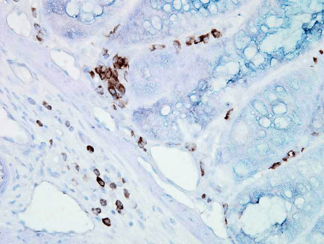
Immunohistochemistry analysis using Mouse Anti-Hsp70 Monoclonal Antibody, Clone BB70 (SMC-106). Tissue: inflamed colon. Species: Mouse. Fixation: Formalin. Primary Antibody: Mouse Anti-Hsp70 Monoclonal Antibody (SMC-106) at 1:10000 for 12 hours at 4°C. Secondary Antibody: Biotin Goat Anti-Mouse at 1:2000 for 1 hour at RT. Counterstain: Mayer Hematoxylin (purple/blue) nuclear stain at 200 µl for 2 minutes at RT. Localization: Inflammatory cells. Magnification: 40x. Inflammatory cells. HSP70/HSC70 stained brown. This image was produced using an amplifying IHC wash buffer. The antibody has therefore been diluted more than is recommended for other applications.
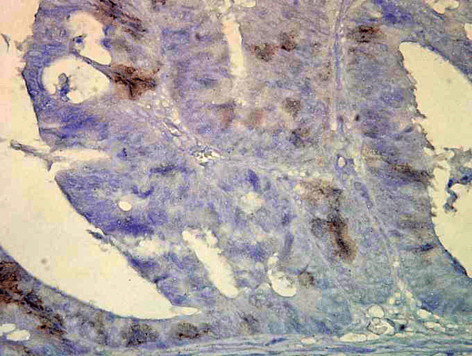
Immunohistochemistry analysis using Mouse Anti-Hsp70 Monoclonal Antibody, Clone BB70 (SMC-106). Tissue: colon carcinoma. Species: Human. Fixation: Formalin. Primary Antibody: Mouse Anti-Hsp70 Monoclonal Antibody (SMC-106) at 1:10000 for 12 hours at 4°C. Secondary Antibody: Biotin Goat Anti-Mouse at 1:2000 for 1 hour at RT. Counterstain: Mayer Hematoxylin (purple/blue) nuclear stain at 200 µl for 2 minutes at RT. Localization: Inflammatory cells. Magnification: 40x. HSP70/HSC70 cells stained brown. This image was produced using an amplifying IHC wash buffer. The antibody has therefore been diluted more than is recommended for other applications.
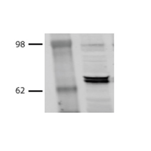
Western Blot analysis of Bovine MDBK cell lysates showing detection of Hsp70 protein using Mouse Anti-Hsp70 Monoclonal Antibody, Clone BB70 (SMC-106). Primary Antibody: Mouse Anti-Hsp70 Monoclonal Antibody (SMC-106) at 1:1000.
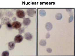
Immunocytochemistry/Immunofluorescence analysis using Mouse Anti-Hsp70 Monoclonal Antibody, Clone BB70 (SMC-106). Tissue: hepatocyte nuclei. Species: Rat. Primary Antibody: Mouse Anti-Hsp70 Monoclonal Antibody (SMC-106) at 1:200. Liver sections were paraffin embedded. First pictures in series show two hours after exposure to stress, the second shows the control.. Courtesy of: G. Matic, University of Belgrade, Serbia.
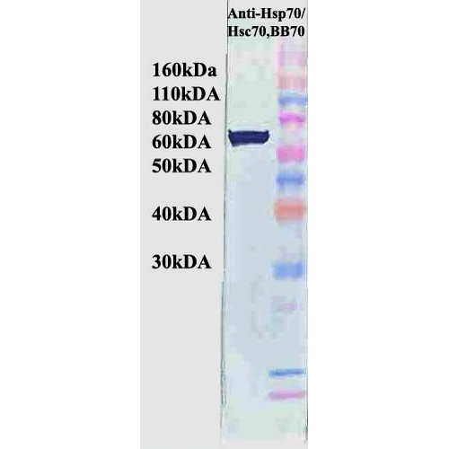
Western Blot analysis of Human Cervical cancer cell line (HeLa) lysate showing detection of Hsp70 protein using Mouse Anti-Hsp70 Monoclonal Antibody, Clone BB70 (SMC-106). Primary Antibody: Mouse Anti-Hsp70 Monoclonal Antibody (SMC-106) at 1:1000. Secondary Antibody: HRP Goat Anti-Rat.
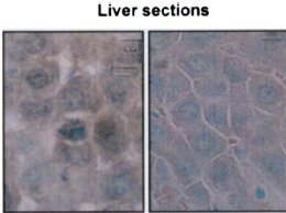
Immunohistochemistry analysis using Mouse Anti-Hsp70 Monoclonal Antibody, Clone BB70 (SMC-106). Tissue: hepatocytes. Species: Rat. Fixation: Paraffin Embedded. Primary Antibody: Mouse Anti-Hsp70 Monoclonal Antibody (SMC-106) at 1:200. Liver sections were paraffin embedded. First pictures in series show two hours after exposure to stress, the second shows the control.. Courtesy of: G. Matic, University of Belgrade, Serbia.
 Powered by Bioz
StressMarq Biosciences :
Based on validation through cited publications.