Properties
| Storage Buffer | PBS pH 7.4, 50% glycerol, 0.09% Sodium azide *Storage buffer may change when conjugated |
| Storage Temperature | -20ºC, Conjugated antibodies should be stored according to the product label |
| Shipping Temperature | Blue Ice or 4ºC |
| Purification | Protein G Purified |
| Clonality | Monoclonal |
| Clone Number | 9E7 |
| Isotype | IgG2a |
| Specificity | Specific for Methylglyoxal modified proteins. Does not detect free Methylglyoxal. Does not cross-react with Acrolein, Hexanoyl Lysine, Malondialdeyhde, 4-Hydroxy-2-hexenal, 4-Hydroxy nonenal, or Crotonaldehyde modified proteins. |
| Cite This Product | StressMarq Biosciences Cat# SMC-516, RRID: AB_2702927 |
| Certificate of Analysis | A 1:1000 dilution of SMC-516 was sufficient for detection of Methylglyoxal in 0.5 µg of Methylglyoxal conjugated to BSA by ECL immunoblot analysis using Goat Anti-Mouse IgG:HRP as the secondary Antibody. |
Biological Description
| Alternative Names | Methylglyoxal Antibody, 2-Oxopropanal Antibody, 2 oxo propanal Antibody, MG Antibody, Pyruvaldehyde Antibody, Methylglyoxal (MG) Antibody, MG-modified protein Antibody |
| Research Areas | Cancer, Lipid peroxidation, Oxidative Stress |
| Scientific Background | Lipid peroxidation occurs when oxidizing agents attack carbon-carbon double bonds found in unsaturated lipids. In addition to membrane degradation, oxidation end-products have been found to damage cell viability through their mutagenic and toxic properties. These downstream functional consequences facilitate the development of disease and premature aging. Methylglyoxal is an alpha-oxoaldehyde generated from lipid peroxidation that reacts with proteins to form adducts(1). It reacts with free amino acids and protein residues to generate advanced glycation endproducts (AGEs) and crosslinks DNA polymerase and substrate DNA to severely inhibit DNA replication (2). |
| References |
1. Negre-Salvayre et al. (2008) Br J Pharmacol. 153(1):6-20. 2. Murata-Kamiya, N., Kamiya, H. (2001) Nucleic Acids Res. 29(16): 3433-3438. |
Product Images
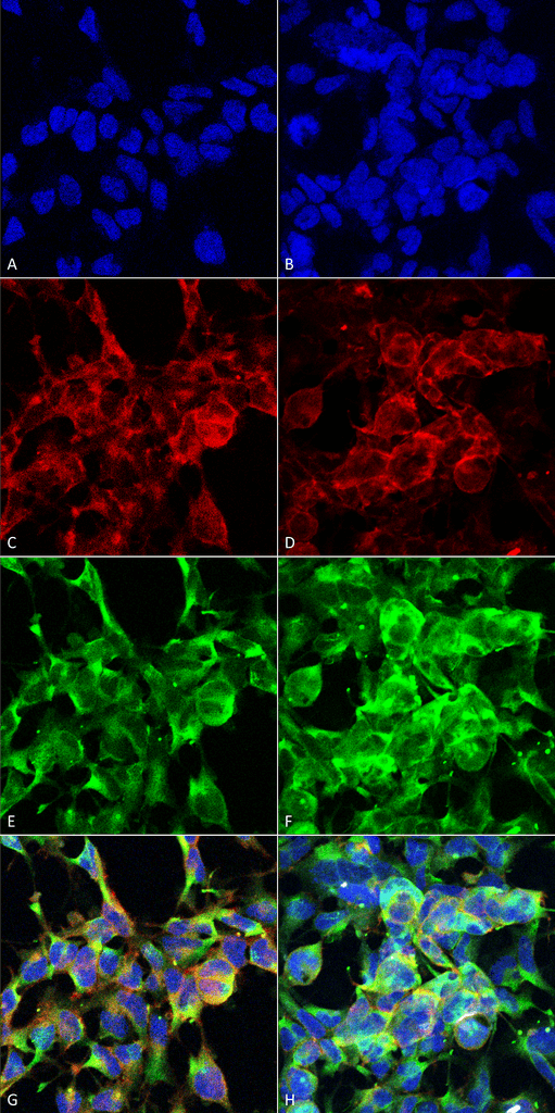
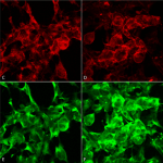
Immunocytochemistry/Immunofluorescence analysis using Mouse Anti-Methylglyoxal Monoclonal Antibody, Clone 9E7 (SMC-516). Tissue: Embryonic kidney epithelial cell line (HEK293). Species: Human. Fixation: 5% Formaldehyde for 5 min. Primary Antibody: Mouse Anti-Methylglyoxal Monoclonal Antibody (SMC-516) at 1:50 for 30-60 min at RT. Secondary Antibody: Goat Anti-Mouse Alexa Fluor 488 at 1:1500 for 30-60 min at RT. Counterstain: Phalloidin Alexa Fluor 633 F-Actin stain; DAPI (blue) nuclear stain at 1:250, 1:50000 for 30-60 min at RT. Magnification: 20X (2X Zoom). (A,C,E,G) – Untreated. (B,D,F,H) – Cells cultured overnight with 50 µM H2O2. (A,B) DAPI (blue) nuclear stain. (C,D) Phalloidin Alexa Fluor 633 F-Actin stain. (E,F) Methylglyoxal Antibody. (G,H) Composite. Courtesy of: Dr. Robert Burke, University of Victoria.
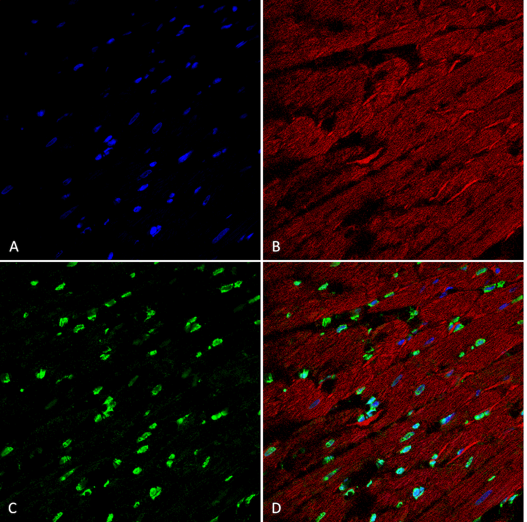
Immunohistochemistry analysis using Mouse Anti-Methylglyoxal Monoclonal Antibody, Clone 9E7 (SMC-516). Tissue: Heart. Species: Rat. Fixation: Formalin fixed, paraffin embedded. Primary Antibody: Mouse Anti-Methylglyoxal Monoclonal Antibody (SMC-516) at 1:25 for 3 hour at RT. Secondary Antibody: Goat Anti-Mouse IgG: Alexa Fluor 488. Counterstain: DAPI (blue) nuclear stain. Magnification: 63X. (A) DAPI (blue) nuclear stain. (B) Actin (C) Methylgyloxal Antibody (D) Composite.
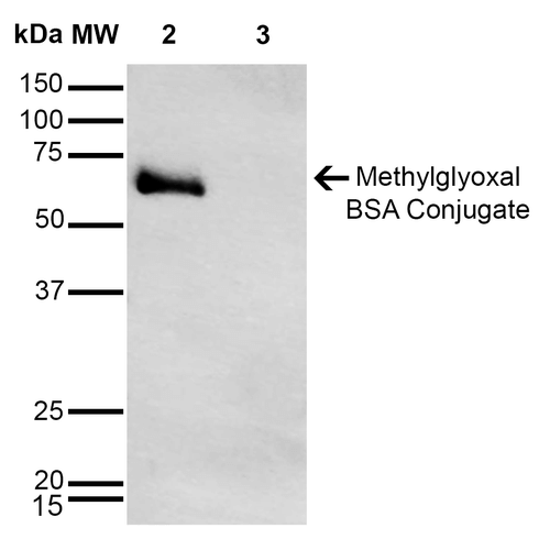
Western Blot analysis of Methylglyoxal-BSA Conjugate showing detection of 67 kDa Methylglyoxal protein using Mouse Anti-Methylglyoxal Monoclonal Antibody, Clone 9E7 (SMC-516). Lane 1: Molecular Weight Ladder (MW). Lane 2: Methylglyoxal-BSA. Lane 3: BSA. Load: 0.5 µg. Block: 5% Skim Milk in TBST. Primary Antibody: Mouse Anti-Methylglyoxal Monoclonal Antibody (SMC-516) at 1:1000 for 2 hours at RT. Secondary Antibody: Goat Anti-Mouse IgG: HRP at 1:1000 for 60 min at RT. Color Development: ECL solution for 5 min in RT. Predicted/Observed Size: 67 kDa.
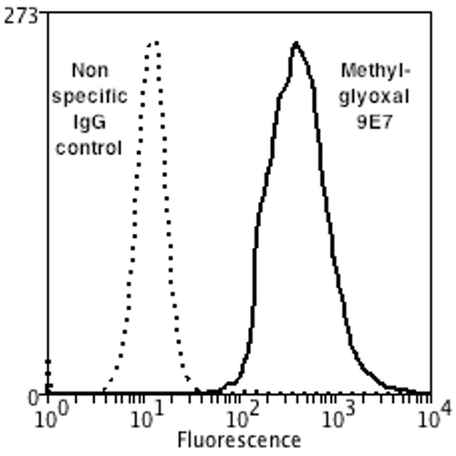
Flow Cytometry analysis using Mouse Anti-Methylglyoxal Monoclonal Antibody, Clone 9E7 (SMC-516). Tissue: Neuroblastoma cells (SH-SY5Y). Species: Human. Fixation: 90% Methanol. Primary Antibody: Mouse Anti-Methylglyoxal Monoclonal Antibody (SMC-516) at 1:50 for 30 min on ice. Secondary Antibody: Goat Anti-Mouse: PE at 1:100 for 20 min at RT. Isotype Control: Non Specific IgG. Cells were subject to oxidative stress by treating with 250 µM H2O2 for 24 hours.

![Mouse Anti-Methylglyoxal Antibody [9E7] used in Flow Cytometry (FCM) on Human Neuroblastoma cells (SH-SY5Y) (SMC-516)](https://www.stressmarq.com/wp-content/uploads/SMC-516_Methylglyoxal_Antibody_9E7_FCM_Human_Neuroblastoma-cells-SH-SY5Y_1-100x100.png)
![Mouse Anti-Methylglyoxal Antibody [9E7] used in Western Blot (WB) on Methylglyoxal-BSA Conjugate (SMC-516)](https://www.stressmarq.com/wp-content/uploads/SMC-516_Methylglyoxal_Antibody_9E7_WB_Methylglyoxal-BSA-Conjugate_1-100x100.png)


StressMarq Biosciences :
Based on validation through cited publications.