Properties
| Storage Buffer | PBS pH7.4, 50% glycerol, 0.09% sodium azide *Storage buffer may change when conjugated |
| Storage Temperature | -20ºC, Conjugated antibodies should be stored according to the product label |
| Shipping Temperature | Blue Ice or 4ºC |
| Purification | Protein G Purified |
| Clonality | Monoclonal |
| Clone Number | L51/82 (Formerly sold as S51-82) |
| Isotype | IgG2a |
| Specificity | Detecs ~92kDa and ~82kDa (pro and active forms). |
| Cite This Product | StressMarq Biosciences Cat# SMC-396, RRID: AB_11232604 |
| Certificate of Analysis | 1 µg/ml of SMC-396 was sufficient for detection of MMP9 in 20 µg of COS-1 cells (lysate) transfected with human MMP9 by colorimetric immunoblot analysis using goat anti-mouse IgG:HRP as the secondary antibody. |
Biological Description
| Alternative Names | MMP-9 Antibody, CLG4B Antibody, 82kDa matrix metalloproteinase-9 Antibody, collagenease type 4 beta Antibody, GELB Antibody, Macrophage gelatinase Antibody, MANDP2 Antibody, Type V collagenase Antibody, 92 kDa gelatinase Antibody, 92 kDa type IV collagenase Antibody, Gelatinase B Antibody, MMP9 Antibody, Matrix metalloproteinase 9 Antibody |
| Research Areas | Adhesion / Extracellular Matrix, Angiogenesis, Cancer, Cardiovascular Growth Factors, Cardiovascular System, Cell Signaling, Enzymes, Extracellular Matrix (ECM), Invasion/microenvironment, Matrix Metalloproteinases, Tumor Biomarker Enzymes, Tumor Biomarkers |
| Cellular Localization | Extracellular Matrix |
| Accession Number | NP_112317.1 |
| Gene ID | 81687 |
| Swiss Prot | P50282 |
| Scientific Background | MMP9, otherwise known as matrix metalloproteinase 9, is involved in the breakdown of extracellular matrix in normal physiological processes such as embryonic development, reproduction and tissue remodeling, as well as in disease processes like arthritis and metastasis (1). Among the family members, MMP-2, MMP-3, MMP-7 and MMP-9 have been characterized as important factors for normal tissue remodeling during embryonic development, wound healing, tumor invasion, angiogenesis, carcinogenesis and apoptosis (2-4). MMP activity correlates with cancer development (2). One mechanism of MMP regulation is transcriptional (5). Once synthesized, MMP exists as a latent proenzyme. Maximum MMP activity requires proteolytic cleavage to generate active MMPs by releasing the inhibitory propeptide domain from the full length protein (5). |
| References |
1. Hirose Y., et al. (2008) Am J Hum Genet. 82(5): 1122-1129. 2. Coussens L.M., et al. (2002) Science 295: 2387-2391. 3. Sternlicht M.D., et al. (1999) Cell 98: 137-146. 4. Vu T.H., et al. (1998) Cell 93: 411-422. 5. Nagase H., et al. (1990) Biochemistry 29: 5783-5789. |
Product Images

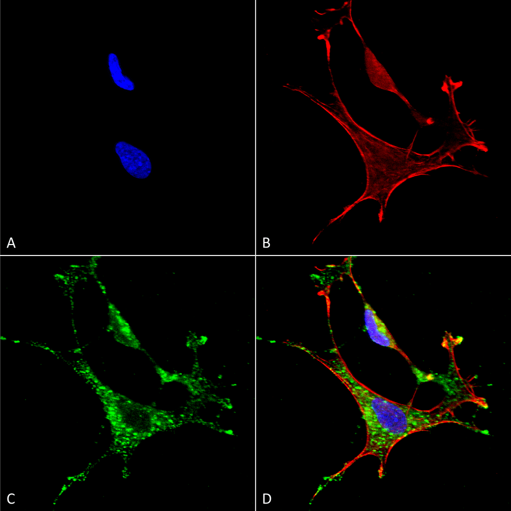
Immunocytochemistry/Immunofluorescence analysis using Mouse Anti-MMP9 Monoclonal Antibody, Clone L51/82 (SMC-396). Tissue: Neuroblastoma cells (SH-SY5Y). Species: Human. Fixation: 4% PFA for 15 min. Primary Antibody: Mouse Anti-MMP9 Monoclonal Antibody (SMC-396) at 1:50 for overnight at 4°C with slow rocking. Secondary Antibody: AlexaFluor 488 at 1:1000 for 1 hour at RT. Counterstain: Phalloidin-iFluor 647 (red) F-Actin stain; Hoechst (blue) nuclear stain at 1:800, 1.6mM for 20 min at RT. (A) Hoechst (blue) nuclear stain. (B) Phalloidin-iFluor 647 (red) F-Actin stain. (C) MMP9 Antibody (D) Composite.

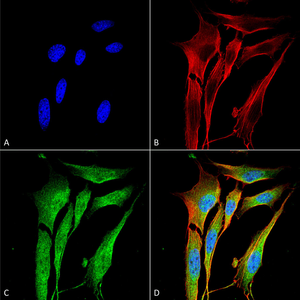
Immunocytochemistry/Immunofluorescence analysis using Mouse Anti-MMP9 Monoclonal Antibody, Clone L51/82 (SMC-396). Tissue: NIH 3T3 (NIH 3T3). Species: Mouse. Fixation: 4% Formaldehyde for 15 min at RT. Primary Antibody: Mouse Anti-MMP9 Monoclonal Antibody (SMC-396) at 1:100 for 60 min at RT. Secondary Antibody: Goat Anti-Mouse ATTO 488 at 1:200 for 60 min at RT. Counterstain: Phalloidin Texas Red F-Actin stain; DAPI (blue) nuclear stain at 1:1000, 1:5000 for 60 min at RT, 5 min at RT. Localization: Cytoplasm . Magnification: 60X. (A) DAPI (blue) nuclear stain. (B) Phalloidin Texas Red F-Actin stain. (C) MMP9 Antibody. (D) Composite.

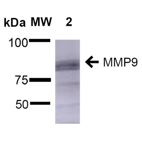
Western Blot analysis of Rat Brain showing detection of ~92 kDa and ~82 kDa (pro and active) MMP9 protein using Mouse Anti-MMP9 Monoclonal Antibody, Clone L51/82 (SMC-396). Lane 1: Molecular Weight Ladder (MW). Lane 2: Rat Brain. Load: 15 µg. Block: 5% Skim Milk in 1X TBST. Primary Antibody: Mouse Anti-MMP9 Monoclonal Antibody (SMC-396) at 1:1000 for 2 hours at RT. Secondary Antibody: Goat Anti-Mouse IgG: HRP at 1:2000 for 60 min at RT. Color Development: ECL solution for 5 min at RT. Predicted/Observed Size: ~92 kDa and ~82 kDa (pro and active).


StressMarq Biosciences :
Based on validation through cited publications.