Properties
| Storage Buffer | PBS, 50% glycerol, 0.09% sodium azide *Storage buffer may change when conjugated |
| Storage Temperature | -20ºC, Conjugated antibodies should be stored according to the product label |
| Shipping Temperature | Blue Ice or 4ºC |
| Purification | Protein G Purified |
| Clonality | Monoclonal |
| Clone Number | 9F12 |
| Isotype | IgG1 |
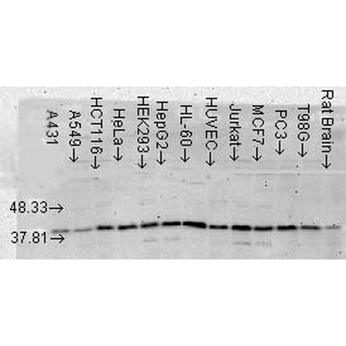
| Specificity | Detects ~38kDa. |
| Cite This Product | StressMarq Biosciences Cat# SMC-152, RRID: AB_2141876 |
| Certificate of Analysis | Detects ~38kDa protein corresponding to p38α MAPK when loaded with 6 ng of purified p38α by chemiluminescent immunoblot analysis using Goat anti-mouse IgG:HRP as the secondary antibody. |
Biological Description
| Alternative Names | CSAID Binding protein 1 Antibody, CSBP1 Antibody, CSBP2 Antibody, EXIP Antibody, MAP kinase MXI2 Antibody, MAPkinase p38alpha Antibody, MAPK14 Antibody, p38 ALPHA Antibody, p38 MAP kinase Antibody, p38 mitogen activated protein kinase Antibody, RK Antibody, SAPK 2A Antibody, Stress activated protein kinase 2A Antibody |
| Research Areas | Cancer, Cell Signaling, Phosphorylation, Post-translational Modifications |
| Cellular Localization | Cytoplasm, Nucleus |
| Accession Number | NP_001306.1 |
| Gene ID | 1432 |
| Swiss Prot | Q16539 |
| Scientific Background | The MAPK (mitogen activated protein kinase) comprises a family of ubiquitous praline-directed, proteinserine/ threonine kinases which signal transduction pathways that control intracellular events including acute responses to hormones and major developmental changes in organisms (1). This super family consists of stress activated protein kinases (SAPKs); extracellular signal-regulated kinases (ERKs); and p38 kinases, each of which forms a separate pathway (2). The kinase members that populate each pathway are sequentially activated by phosphorylation. Upon activation, p38 MAPK/SAPK2α translocates into the nucleus where it phosphorylates one or more nuclear substrates, effecting transcriptional changes and other cellular processes involved in cell growth, division, differentiation, inflammation, and death (3). Specifically p38 always acts as a pro-apoptotic factor with its activation leading to the release of cytochrome c from mitochondria and cleavage of caspase 3 and its downstream effector, PARP (4). p38 MAPK is activated by a variety of chemical stress inducers including hydrogen peroxide, heavy metals, anisomycin, sodium salicylate, LPS, and biological stress signals such as tumor necrosis factor, interleukin-1, ionizing and UV irradiation, hyperosmotic stress and chemotherapeutic drugs (5). As a result, p38 alpha has been widely validated as a target for inflammatory disease including rheumatoid arthritis, COPD and psoriasis (6) and has also been implicated in cancer, CNS and diabetes (7). |
| References |
1. Pearson, G. et al (2001). Endocrine Reviews 22 (2): 153-183. 2. Fan, Y. et al (2007) Mol. Cells 23 (1): 30-38. 3. Han, J. et al. (1994) Science 265: 808-811. 4. Van, L. A., et al. (2004) Faseb J. 18: 1946−1948. 5. Deng et al. (2003) Cell. 115: 61-70. 6. Salojin KV, et al. (2006) J Immunol. 176 (3):1899-907. 7. Medicherla S. et al. (2006). J Pharmacol Exp Ther.318(1): 99-107. |
Product Images

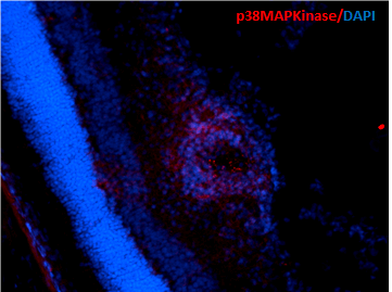
Immunohistochemistry analysis using Mouse Anti-p38 MAPK Monoclonal Antibody, Clone 9F12 (SMC-152). Tissue: Retinal Injury Model. Species: Mouse. Primary Antibody: Mouse Anti-p38 MAPK Monoclonal Antibody (SMC-152) at 1:1000. Secondary Antibody: Alexa Fluor 594 Goat Anti-Mouse (red). Courtesy of: Dr. Rajashekhar Gangaraju, University of Indiana, Department of Ophthalmology, Eugene and Marilyn Glick Eye Institute.

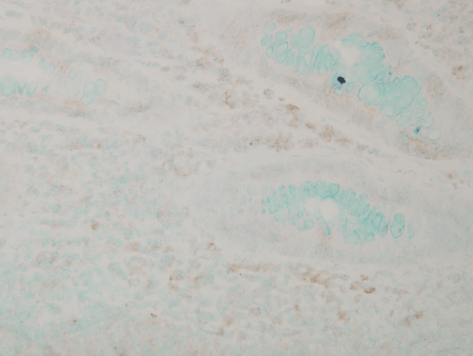
Immunohistochemistry analysis using Mouse Anti-p38 MAPK Monoclonal Antibody, Clone 9F12 (SMC-152). Tissue: colon carcinoma. Species: Human. Fixation: Formalin. Primary Antibody: Mouse Anti-p38 MAPK Monoclonal Antibody (SMC-152) at 1:10000 for 12 hours at 4°C. Secondary Antibody: Biotin Goat Anti-Mouse at 1:2000 for 1 hour at RT. Counterstain: Mayer Hematoxylin (purple/blue) nuclear stain at 200 µl for 2 minutes at RT. Magnification: 40x.

Western Blot analysis of Human Cell lysates showing detection of p38 MAPK protein using Mouse Anti-p38 MAPK Monoclonal Antibody, Clone 9F12 (SMC-152). Load: 15 µg. Block: 1.5% BSA for 30 minutes at RT. Primary Antibody: Mouse Anti-p38 MAPK Monoclonal Antibody (SMC-152) at 1:1000 for 2 hours at RT. Secondary Antibody: Sheep Anti-Mouse IgG: HRP for 1 hour at RT.


![Mouse Anti-p38 MAPK Antibody [9F12] used in Immunohistochemistry (IHC) on Human colon carcinoma (SMC-152)](https://www.stressmarq.com/wp-content/uploads/SMC-152_p38-MAPK_Antibody_9F12_IHC_Human_colon-carcinoma_40x_1-100x100.png)
![Mouse Anti-p38 MAPK Antibody [9F12] used in Western Blot (WB) on Human Cell lysates (SMC-152)](https://www.stressmarq.com/wp-content/uploads/SMC-152_p38-MAPK_Antibody_9F12_WB_Human_Cell-lysates_1-100x100.png)


StressMarq Biosciences :
Based on validation through cited publications.