Properties
| Storage Buffer | PBS pH7.4, 50% glycerol, 0.09% sodium azide *Storage buffer may change when conjugated |
| Storage Temperature | -20ºC, Conjugated antibodies should be stored according to the product label |
| Shipping Temperature | Blue Ice or 4ºC |
| Purification | Protein G Purified |
| Clonality | Monoclonal |
| Clone Number | N23b/49 (Formerly sold as S23b-49) |
| Isotype | IgG1 |
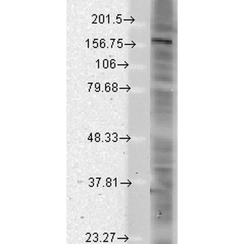
| Specificity | Detects ~160kDa. Recognizes Shank1, 2 and 3. |
| Cite This Product | StressMarq Biosciences Cat# SMC-327, RRID: AB_2301756 |
| Certificate of Analysis | 1 µg/ml of SMC-327 was sufficient for detection of Shank1-4 in 10 µg of rat brain lysate by colorimetric immunoblot analysis using Goat anti-mouse IgG:HRP as the secondary antibody. |
Biological Description
| Alternative Names | Cortactin binding protein 1 Antibody, Cortactin SH3 domain-binding protein Antibody, Cortactin-binding protein 1 Antibody, CortBP1 Antibody, CTTNBP1 Antibody, GKAP/SAPAP interacting protein Antibody, GKAP/SAPAP-interacting protein Antibody, KIAA1022 Antibody, Proline rich synapse associated protein 1 Antibody, Proline-rich synapse-associated protein 1 Antibody, ProSAP1 Antibody, SH3 and multiple ankyrin repeat domains protein 2 Antibody, SHAN2_RAT Antibody, SHANK Antibody, Shank2 Antibody, SPANK-3 Antibody |
| Research Areas | Cell Markers, Cell Signaling, Neuron Markers, Neuroscience, Postsynaptic Markers, Scaffold Proteins |
| Cellular Localization | Cell Junction, Cell projection, Cytoplasm, Growth cone, Postsynaptic cell membrane, Postsynaptic density, Synapse |
| Gene ID | 171093 |
| Swiss Prot | Q9QX74 |
| Scientific Background | Shank proteins make up a family of scaffold proteins identified through their interaction with a variety of membrane and cytoplasmic proteins (1). Shank proteins at postsynaptic sites of excitatory synapses play roles in signal transmission into the postsynaptic neuron. Studies suggest that Shank2 is expressed in the neurons of the developing retina, and could play a role in the neuronal differentiation of the developing retina (2). Other recent studies suggest that the disruption of glutamate receptors at the Shank postsynaptic platform could contribute to the destruction of the postsynaptic density, which underlies the synaptic dysfunction and loss in Alzheimer's disease (3). |
| References |
1. Sheng M., and Kim E. (2000) Journal of Cell Science. 113: 1851-1856. 2. Kim J.H., et al. (2009) Exp Mol Med. 41(4): 236-242. 3. Gong Y., et al. (2009) Brain Res. 1292: 191-198. |
Product Images

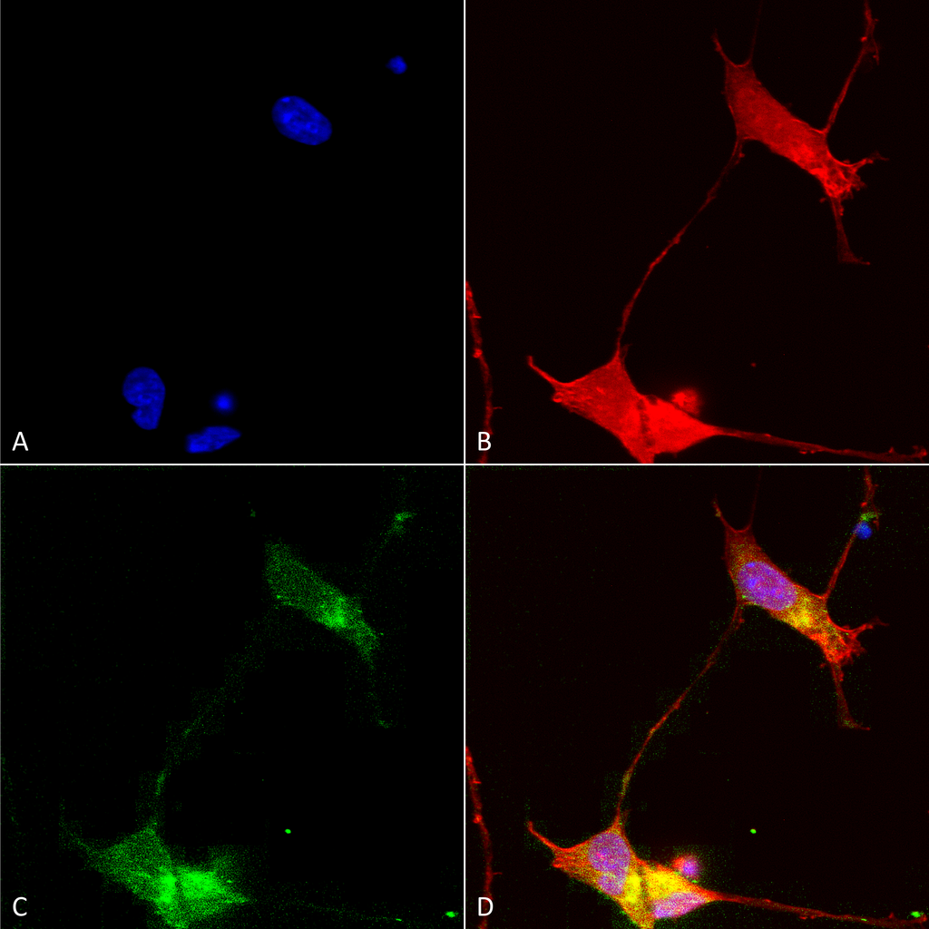
Immunocytochemistry/Immunofluorescence analysis using Mouse Anti-SHANK (pan) Monoclonal Antibody, Clone S23b-49 (SMC-327). Tissue: Neuroblastoma cells (SH-SY5Y). Species: Human. Fixation: 4% PFA for 15 min. Primary Antibody: Mouse Anti-SHANK (pan) Monoclonal Antibody (SMC-327) at 1:50 for overnight at 4°C with slow rocking. Secondary Antibody: AlexaFluor 488 at 1:1000 for 1 hour at RT. Counterstain: Phalloidin-iFluor 647 (red) F-Actin stain; Hoechst (blue) nuclear stain at 1:800, 1.6mM for 20 min at RT. (A) Hoechst (blue) nuclear stain. (B) Phalloidin-iFluor 647 (red) F-Actin stain. (C) SHANK (pan) Antibody (D) Composite.

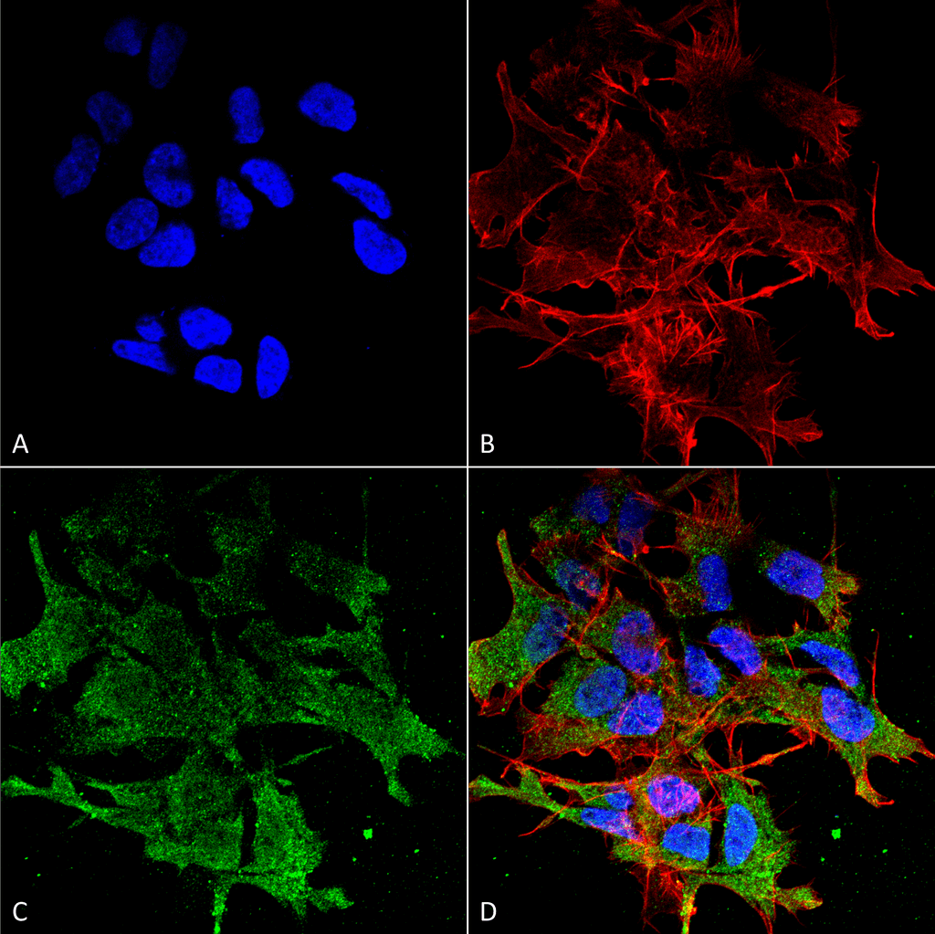
Immunocytochemistry/Immunofluorescence analysis using Mouse Anti-SHANK (pan) Monoclonal Antibody, Clone S23b-49 (SMC-327). Tissue: Neuroblastoma cell line (SK-N-BE). Species: Human. Fixation: 4% Formaldehyde for 15 min at RT. Primary Antibody: Mouse Anti-SHANK (pan) Monoclonal Antibody (SMC-327) at 1:100 for 60 min at RT. Secondary Antibody: Goat Anti-Mouse ATTO 488 at 1:200 for 60 min at RT. Counterstain: Phalloidin Texas Red F-Actin stain; DAPI (blue) nuclear stain at 1:1000, 1:5000 for 60 min at RT, 5 min at RT. Localization: Cytoplasm . Magnification: 60X. (A) DAPI (blue) nuclear stain. (B) Phalloidin Texas Red F-Actin stain. (C) SHANK (pan) Antibody. (D) Composite.

Western Blot analysis of Rat brain membrane lysate showing detection of SHANK protein using Mouse Anti-SHANK Monoclonal Antibody, Clone S23b-49 (SMC-327). Load: 15 µg. Block: 1.5% BSA for 30 minutes at RT. Primary Antibody: Mouse Anti-SHANK Monoclonal Antibody (SMC-327) at 1:1000 for 2 hours at RT. Secondary Antibody: Sheep Anti-Mouse IgG: HRP for 1 hour at RT.


StressMarq Biosciences :
Based on validation through cited publications.