Properties
| Storage Buffer | PBS pH7.4, 50% glycerol, 0.09% sodium azide *Storage buffer may change when conjugated |
| Storage Temperature | -20ºC, Conjugated antibodies should be stored according to the product label |
| Shipping Temperature | Blue Ice or 4ºC |
| Purification | Protein G Purified |
| Clonality | Monoclonal |
| Clone Number | N104/32 (Formerly sold as S104-32) |
| Isotype | IgG1 |
| Specificity | Detects ~50kDa. |
| Cite This Product | StressMarq Biosciences Cat# SMC-401, RRID: AB_11232603 |
| Certificate of Analysis | 1 µg/ml of SMC-401 was sufficient for detection of SNAT1 in 20 µg of lysates from neocortical neurons cultured under amino acid starvation conditions and assayed by colorimetric immunoblot analysis using goat anti-mouse IgG:HRP as the secondary antibody. |
Biological Description
| Alternative Names | SLC38A1 Antibody, amino acids transporter A1 Antibody, ATA1 Antibody, NAT2 Antibody, sodium coupled neutral amino acids transporter 1 Antibody, Amino acid transporter system A1 Antibody, N-system amino acid transporter 2 Antibody, S38A1 Antibody, SAT1 Antibody, SNAT 1 Antibody, Solute carrier family 38 member 1 Antibody, System A amino acid transporter 1 Antibody, System N amino acid transporter 1 Antibody |
| Research Areas | Cell Signaling, Neuroscience, Neurotransmitter Transporters, Protein Trafficking, Pumps/Transporters |
| Cellular Localization | Cell membrane |
| Accession Number | NP_620187.1 |
| Gene ID | 170567 |
| Swiss Prot | Q9JM15 |
| Scientific Background | The sodium-coupled neutral amino acid transporters (SNAT) of the SLC38 gene family include System A subtypes SNAT1, SNAT2 and SNAT4 and System N subtypes SNAT3 and SNAT5. The SLC38 transporters are essential for the uptake of nutrients, energy production, metabolism, detoxification, and the cycling of neurotransmitters. The SNAT1 protein, also designated ATA1 or NAT2 is encoded by the human gene SLC38A1 which maps to chromosome 12q13.11. SNAT1 is responsible for the transport of glutamine, an intermediate in the synthesis of urea, and may be involved in the generation of glutamate in the retina. SNAT1 protein may be detected in some tissues such as heart, brain and placenta and expression levels are enriched in certain neuronal populations within the CNS. SNAT1 is not present in astrocytes. |
| References |
1. Hatanaka T., et al. ( 2000) Biochim. Biophys. Acta 1467: 1-6. 2. Wang H., et al. (2000) Biochem. Biophys. Res. Commun. 273: 1175-1179. 3. Gu S., et al. (2001) J. Biol. Chem. 276: 24137-24144. 4. Freeman T.L., et al. (2002) Arch. Biochem. Biophys. 400: 215-222. 5. Palii S.S., et al. (2004) J. Biol. Chem. 279: 3463-3471. 6. Sidoryk M., et al. (2004) Neuroreport. 15: 575-578. Erratum in: 2004. Neuroreport. 15: 533. 7. Desforges,M., et al. (2006) Am. J. Physiol. Cell. Physiol. 290: 305-312. |
Product Images

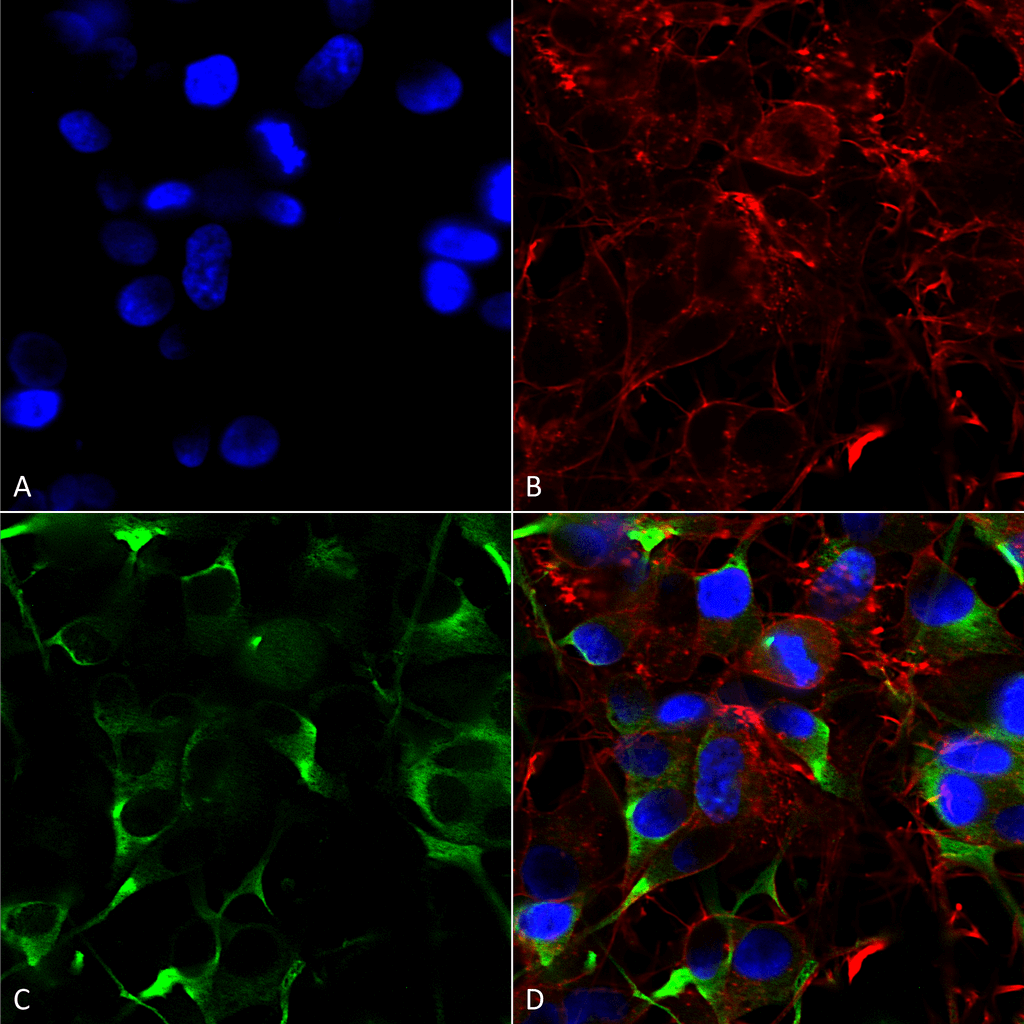
Immunocytochemistry/Immunofluorescence analysis using Mouse Anti-SNAT1 Monoclonal Antibody, Clone N104/32 (SMC-401). Tissue: Neuroblastoma cells (SH-SY5Y). Species: Human. Fixation: 4% PFA for 15 min. Primary Antibody: Mouse Anti-SNAT1 Monoclonal Antibody (SMC-401) at 1:200 for overnight at 4°C with slow rocking. Secondary Antibody: AlexaFluor 488 at 1:1000 for 1 hour at RT. Counterstain: Phalloidin-iFluor 647 (red) F-Actin stain; Hoechst (blue) nuclear stain at 1:800, 1.6mM for 20 min at RT. (A) Hoechst (blue) nuclear stain. (B) Phalloidin-iFluor 647 (red) F-Actin stain. (C) SNAT1 Antibody (D) Composite.

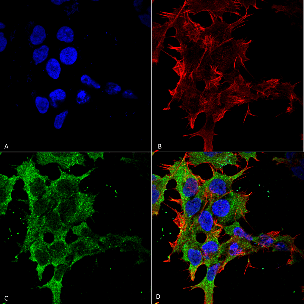
Immunocytochemistry/Immunofluorescence analysis using Mouse Anti-SNAT1 Monoclonal Antibody, Clone N104/32 (SMC-401). Tissue: Neuroblastoma cell line (SK-N-BE). Species: Human. Fixation: 4% Formaldehyde for 15 min at RT. Primary Antibody: Mouse Anti-SNAT1 Monoclonal Antibody (SMC-401) at 1:100 for 60 min at RT. Secondary Antibody: Goat Anti-Mouse ATTO 488 at 1:100 for 60 min at RT. Counterstain: Phalloidin Texas Red F-Actin stain; DAPI (blue) nuclear stain at 1:1000, 1:5000 for 60min RT, 5min RT. Localization: Cell Membrane. Magnification: 60X. (A) DAPI (blue) nuclear stain. (B) Phalloidin Texas Red F-Actin stain. (C) SNAT1 Antibody. (D) Composite.


![Mouse Anti-SLC38A1 Antibody [N104/32] used in Western Blot (WB) on Rat brain membrane lysate (SMC-401)](https://www.stressmarq.com/wp-content/uploads/SMC-401_SLC38A1_Antibody_N104-32_WB_Rat_brain-membrane-lysate_1.png)


Mehrdad Khajavi :
We used this antibody for both western blot and also used to stain corneal tissue and HMVECs for IF by confocal microscopy. It worked great.