Properties
| Storage Buffer | PBS pH 7.4, 50% glycerol, 0.1% sodium azide *Storage buffer may change when conjugated |
| Storage Temperature | -20ºC, Conjugated antibodies should be stored according to the product label |
| Shipping Temperature | Blue Ice or 4ºC |
| Purification | Protein G Purified |
| Clonality | Monoclonal |
| Clone Number | N374/48 (Formerly sold as S374-48) |
| Isotype | IgG2b |
| Specificity | Detects ~50kDa. Does not cross-react with TASK3. |
| Cite This Product | StressMarq Biosciences Cat# SMC-473, RRID: AB_2702225 |
| Certificate of Analysis | 1 µg/ml of SMC-473 was sufficient for detection of TASK1 Potassium Channel in 20 µg of rat brain lysate by colorimetric immunoblot analysis using Goat anti-mouse IgG:HRP as the secondary antibody. |
Biological Description
| Alternative Names | Potassium channel subfamily K member 3 Antibody, KCNK3 Antibody, Acid sensitive potassium channel protein TASK 1 Antibody, Cardiac two pore background K(+) channel Antibody, cTBAK 1 Antibody, K2p3.1 Antibody, KCNK9 Antibody, OAT1 Antibody, potassium channel subfamily K member 3 Antibody, rTASK Antibody, TASK 1 Antibody, TBAK1 Antibody, TWIK related acid sensitive K+ channel Antibody, Two pore potassium channel KT3.1 Antibody, Two pore K(+) channel KT3.1 Antibody |
| Research Areas | Ion Channels, Neuroscience, Potassium Channels, Tandem Pore Domain Potassium Channels |
| Cellular Localization | Membrane |
| Accession Number | NP_203694.1 |
| Gene ID | 29553 |
| Swiss Prot | O54912 |
| Scientific Background | K+ channels are divided into three subclasses reflecting the number of transmembrane segments (TMS), which are designated 6TMS, 4TMS and 2TMS. Members of the 4TMS class contain two distinct pore regions and include TWIK, TREK, TRAAK and TASK. TASK channels are highly sensitive to external pH in the physiological range. TASK-1 is expressed in brain and in rat heart, with high levels of expression in the right atrium. TASK-2, mainly expressed in kidney, is localized in cortical distal tubules and collecting ducts, suggesting a role in renal K+ transport. TASK-3 from rat cerebellum shares 54% identity with TASK-1, but less than 30% identity with TASK-2 and other tandem pore K+ channels. |
Product Images

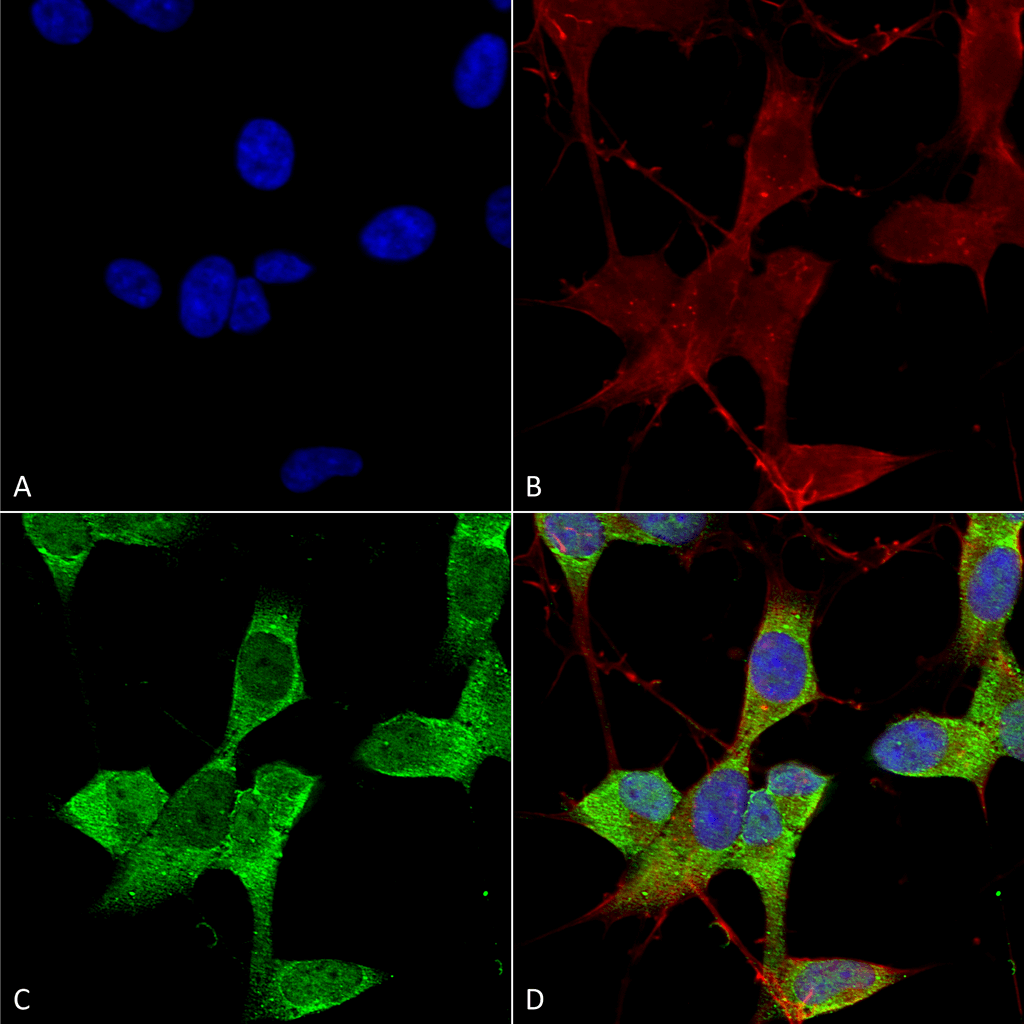
Immunocytochemistry/Immunofluorescence analysis using Mouse Anti-TASK1 Potassium Channel Monoclonal Antibody, Clone N374/48 (SMC-473). Tissue: Neuroblastoma cells (SH-SY5Y). Species: Human. Fixation: 4% PFA for 15 min. Primary Antibody: Mouse Anti-TASK1 Potassium Channel Monoclonal Antibody (SMC-473) at 1:50 for overnight at 4°C with slow rocking. Secondary Antibody: AlexaFluor 488 at 1:1000 for 1 hour at RT. Counterstain: Phalloidin-iFluor 647 (red) F-Actin stain; Hoechst (blue) nuclear stain at 1:800, 1.6mM for 20 min at RT. (A) Hoechst (blue) nuclear stain. (B) Phalloidin-iFluor 647 (red) F-Actin stain. (C) TASK1 Potassium Channel Antibody (D) Composite.

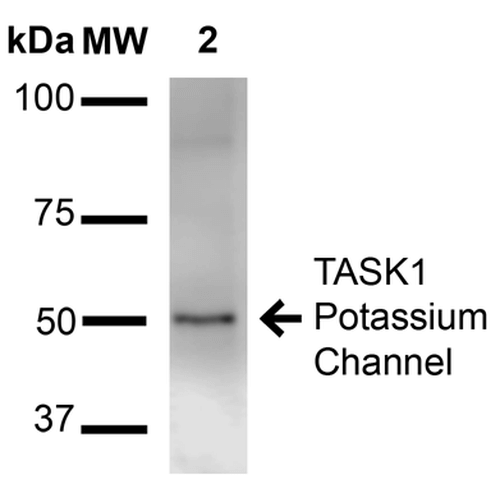
Western Blot analysis of Rat Brain Membrane showing detection of ~50 kDa TASK1 Potassium Channel protein using Mouse Anti-TASK1 Potassium Channel Monoclonal Antibody, Clone N374/48 (SMC-473). Lane 1: Molecular Weight Ladder (MW). Lane 2: Rat brain membrane. Load: 15 µg. Block: 2% BSA and 2% Skim Milk in 1X TBST. Primary Antibody: Mouse Anti-TASK1 Potassium Channel Monoclonal Antibody (SMC-473) at 1:1000 for 16 hours at 4°C. Secondary Antibody: Goat Anti-Mouse IgG: HRP at 1:2000 for 60 min at RT. Color Development: ECL solution for 6 min at RT. Predicted/Observed Size: ~50 kDa.

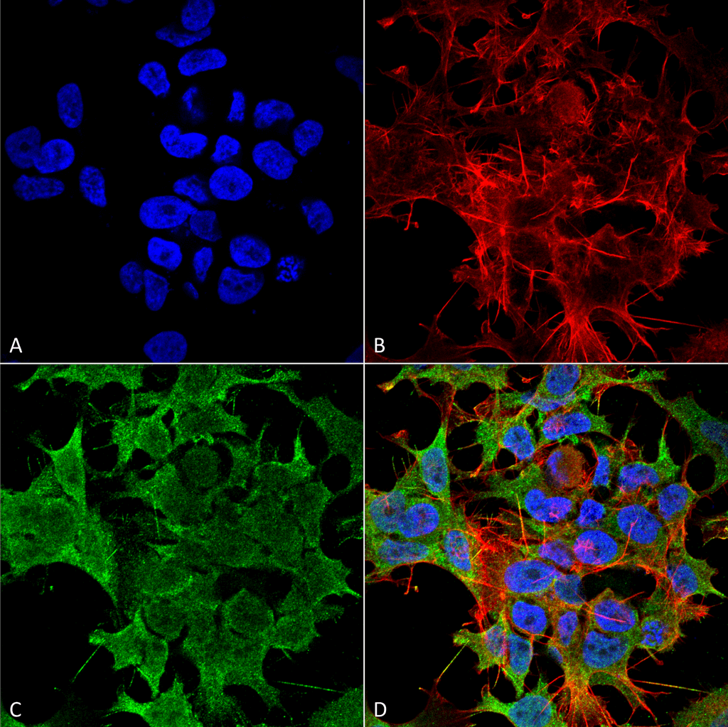
Immunocytochemistry/Immunofluorescence analysis using Mouse Anti-TASK1 Potassium Channel Monoclonal Antibody, Clone N374/48 (SMC-473). Tissue: Neuroblastoma cell line (SK-N-BE). Species: Human. Fixation: 4% Formaldehyde for 15 min at RT. Primary Antibody: Mouse Anti-TASK1 Potassium Channel Monoclonal Antibody (SMC-473) at 1:100 for 60 min at RT. Secondary Antibody: Goat Anti-Mouse ATTO 488 at 1:100 for 60 min at RT. Counterstain: Phalloidin Texas Red F-Actin stain; DAPI (blue) nuclear stain at 1:1000, 1:5000 for 60min RT, 5min RT. Localization: Membrane. Magnification: 60X. (A) DAPI (blue) nuclear stain. (B) Phalloidin Texas Red F-Actin stain. (C) TASK1 Potassium Channel Antibody. (D) Composite.


Reviews
There are no reviews yet.