Properties
| Storage Buffer | PBS pH 7.4, 50% glycerol, 0.09% Sodium azide *Storage buffer may change when conjugated |
| Storage Temperature | -20ºC, Conjugated antibodies should be stored according to the product label |
| Shipping Temperature | Blue Ice or 4ºC |
| Purification | Protein G Purified |
| Clonality | Monoclonal |
| Clone Number | 7E4 |
| Isotype | IgG2a |
| Specificity | Detects ~92 kDa. |
| Cite This Product | Mouse Anti-Human VPS35 Monoclonal (StressMarq Biosciences, Victoria BC, Cat# SMC-602) |
| Certificate of Analysis | A 1:1000 dilution of SMC-602 was sufficient for detection of VPS35 in 10 µg of SH-SY5Y by ECL immunoblot analysis using Goat Anti-Mouse IgG:HRP as the secondary antibody. |
Biological Description
| Alternative Names | Vacuolar protein sorting-associated protein 35, MEM3, PARK17, VPS35 retromer complex component, maternal-embryonic 3, vesicle protein sortin 35, TCCCTA00141, FLJ10752 |
| Research Areas | Alzheimer's Disease, Cell Signaling, Golgi Proteins, Membrane Trafficking Proteins, Neurodegeneration, Neuroscience, Parkinson's Disease, Protein Trafficking |
| Cellular Localization | Cytoplasm, Endosome, Lysosome, Membrane, Vesicles |
| Accession Number | NP_060676.2 |
| Gene ID | 55737 |
| Swiss Prot | Q96QK1 |
| Scientific Background | Vacuolar Protein Sorter-35 (VPS35) is a component of the retromer complex, which is essential for endosome-to-Golgi retrieval of membrane proteins. VPS35 mutations such as D620N have been linked to Parkinson’s Disease (PD) (1,2) and affect retromer function, protein homeostasis, and mitochondria (3). |
| References |
1. Vilarino-Guell, C. et al. (2011) Am J Hum Genet 89:162–167 2. Zimprich, A. et al. (2011) Am J Hum Genet 89:168–175 3. Rahman, A.A., Morrison, B.E. (2019) Neurosci 401:1-10. |
Product Images
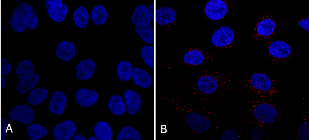
Immunocytochemistry/Immunofluorescence analysis using Mouse Anti-VPS35 Monoclonal Antibody, Clone 7E4 (SMC-602). Tissue: A549 cells. Species: Human. Primary Antibody: Mouse Anti-VPS35 Monoclonal Antibody (SMC-602) at 1:5 (tissue culture supernatant). Secondary Antibody: Donkey anti-mouse: Alexa Fluor 594 at 1:4000 in 0.2% BSA PBS. Counterstain: DAPI. Localization: Vesicles. A) VPS35 KO A549 cells B) WT A549 cells. Courtesy of: Dario Alessi Lab, University of Dundee.
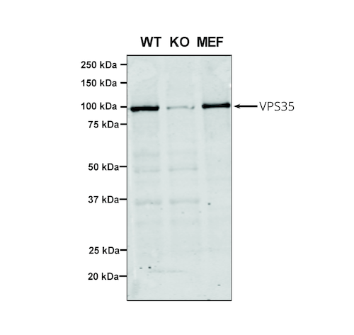
Western Blot analysis of Human, Mouse A549, MEF showing detection of VPS35 protein using Mouse Anti-VPS35 Monoclonal Antibody, Clone 7E4 (SMC-602). Lane 1: Molecular Weight Ladder. Lane 2: VPS35 KO A549 cells. Lane 3: mouse embryonic fibroblast cells.. Load: 8 µg each A549 and MEF. Primary Antibody: Mouse Anti-VPS35 Monoclonal Antibody (SMC-602) at 1:5 (tissue culture supernatant). Secondary Antibody: Donkey anti-mouse IRDye 800CW at 1:25000 in TBS-T.
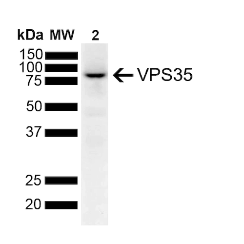
Western Blot analysis of Human SH-SY5Y lysates showing detection of 91.7 kDa VPS35 protein using Mouse Anti-VPS35 Monoclonal Antibody, Clone 7E4 (SMC-602). Lane 1: Molecular Weight Ladder. Lane 2: SH-SY5Y. Load: 10 μg. Block: 5% Skim Milk powder in TBST. Primary Antibody: Mouse Anti-VPS35 Monoclonal Antibody (SMC-602) at 1:1000 for 2 hours at RT with shaking. Secondary Antibody: Goat anti-mouse IgG:HRP at 1:4000 for 1 hour at RT with shaking. Color Development: Chemiluminescent for HRP (Moss) for 5 min in RT. Predicted/Observed Size: 91.7 kDa.
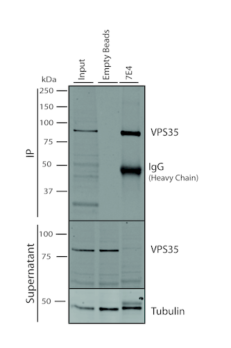
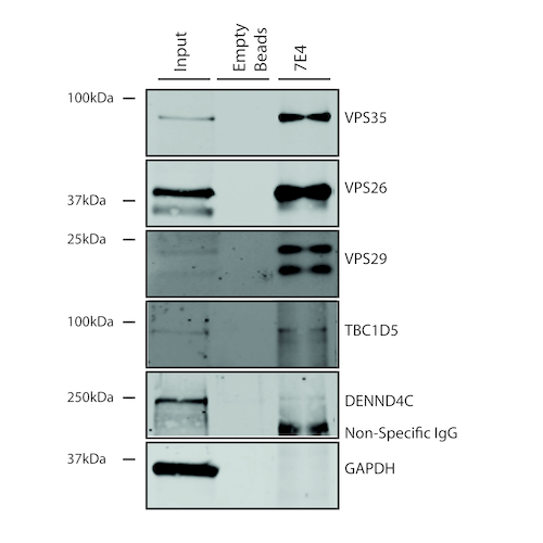
Immunoprecipitation analysis using Mouse Anti-VPS35 Monoclonal Antibody, Clone 7E4 (SMC-602). Tissue: A549 cells. Species: Human. Primary Antibody: Mouse Anti-VPS35 Monoclonal Antibody (SMC-602). 500 µL cell culture supernatants were incubated with 10 µL of Protein A/G resin beads for 1 hour at 4⁰C. SMC-602 clone 7E4 depletes virtually all of the VPS35 from the A549 cell extract..
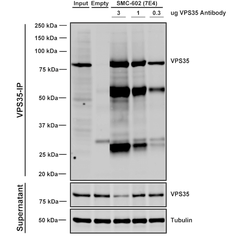
Immunoprecipitation analysis using Mouse Anti-VPS35 Monoclonal Antibody, Clone 7E4 (SMC-602). Tissue: embryonic fibroblast. Species: Mouse. Primary Antibody: Mouse Anti-VPS35 Monoclonal Antibody (SMC-602). Three amounts of SMC-602 (3, 1 and 0.3 ug) were non-covalently coupled to 10uL of A/G sepharose beads for 1 hour at 4°C and next incubated with 250ug of MEF lysate for 2 hours at 4°C.
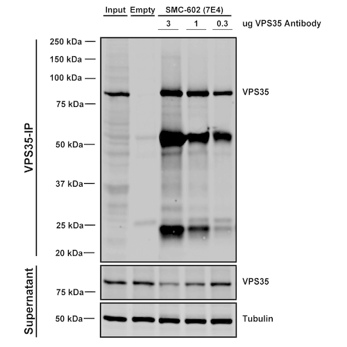
Immunoprecipitation analysis using Mouse Anti-VPS35 Monoclonal Antibody, Clone 7E4 (SMC-602). Tissue: A549 cells. Species: Human. Primary Antibody: Mouse Anti-VPS35 Monoclonal Antibody (SMC-602). Three amounts of SMC-602 (3, 1 and 0.3 ug) were non-covalently coupled to 10uL of A/G sepharose beads for 1 hour at 4°C and next incubated with 250ug of A549 lysate for 2 hours at 4°C.
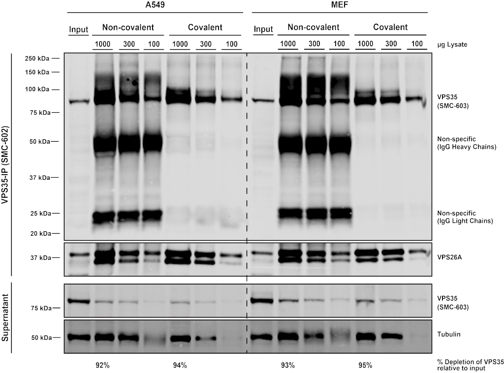
Immunoprecipitation analysis using Mouse Anti-VPS35 Monoclonal Antibody, Clone 7E4 (SMC-602). Tissue: MEF, A549 cells. Species: Human, Mouse. Primary Antibody: Mouse Anti-VPS35 Monoclonal Antibody (SMC-602) at 1:5 (tissue culture supernatant). 10 ug antibody were coupled to 10 uL A/G resin beads either covalently (with DMP) or non-covalently (1 hour at 4 degrees). The antibody immunoprecipitates VPS35 in mouse and human cells effectively when covalently coupled to the beads..

![Mouse Anti-VPS35 Antibody [7E4] used in Immunoprecipitation (IP) on Human A549 cells (SMC-602)](https://www.stressmarq.com/wp-content/uploads/SMC-602_VPS35_Antibody_7E4_IP_Human_A549-cells_1-100x100.png)
![Mouse Anti-VPS35 Antibody [7E4] used in Immunoprecipitation (IP) on Human A549 cells (SMC-602)](https://www.stressmarq.com/wp-content/uploads/SMC-602_VPS35_Antibody_7E4_IP_Human_A549-cells_2-100x100.png)
![Mouse Anti-VPS35 Antibody [7E4] used in Western Blot (WB) on Human, Mouse A549, MEF (SMC-602)](https://www.stressmarq.com/wp-content/uploads/SMC-602_VPS35_Antibody_7E4_WB_Human-Mouse_A549-MEF_1-100x100.png)
![Mouse Anti-VPS35 Antibody [7E4] used in Western Blot (WB) on Human SH-SY5Y lysates (SMC-602)](https://www.stressmarq.com/wp-content/uploads/SMC-602_VPS35_Antibody_7E4_WB_Human_SH-SY5Y-lysates_2-100x100.png)
![Mouse Anti-VPS35 Antibody [7E4] used in Immunoprecipitation (IP) on Human A549 cells (SMC-602)](https://www.stressmarq.com/wp-content/uploads/SMC-602_VPS35_Antibody_7E4_IP_Human_A549-cells_3-100x100.png)
![Mouse Anti-VPS35 Antibody [7E4] used in Immunoprecipitation (IP) on Mouse embryonic fibroblast (SMC-602)](https://www.stressmarq.com/wp-content/uploads/SMC-602_VPS35_Antibody_7E4_IP_Mouse_embryonic-fibroblast_4-100x100.png)
![Mouse Anti-VPS35 Antibody [7E4] used in Immunoprecipitation (IP) on Human, Mouse MEF, A549 cells (SMC-602)](https://www.stressmarq.com/wp-content/uploads/SMC-602_VPS35_Antibody_7E4_IP_Human-Mouse_MEF-A549-cells_5-100x100.png)


Reviews
There are no reviews yet.