Properties
| Storage Buffer | PBS pH 7.4, 50% glycerol, 0.09% sodium azide *Storage buffer may change when conjugated |
| Storage Temperature | -20ºC, Conjugated antibodies should be stored according to the product label |
| Shipping Temperature | Blue Ice or 4ºC |
| Purification | Protein A purified |
| Clonality | Polyclonal |
| Specificity | Detects monomeric and homodimeric GDNF |
| Cite This Product | Rabbit Anti-Human GDNF Polyclonal (StressMarq Biosciences, Victoria BC, Cat# SPC-810) |
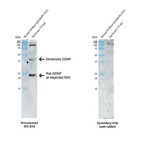
| Certificate of Analysis | 1 µg/mL of SPC-810 was sufficient for detection of GDNF in 15 µg of rat brain tissue lysate by colorimetric immunoblot analysis using goat anti-rabbit IgG:HRP as the secondary antibody. |
Biological Description
| Alternative Names | Glial cell line-derived neurotrophic factor Antibody, Astrocyte-derived trophic factor Antibody, Astrocyte derived trophic factor 1 Antibody, Glial cell derived neurotrophic factor Antibody, ATF 1 Antibody, ATF 2 Antibody, Glial Cell Line Derived Neurotrophic Factor Antibody, ATF Antibody, Atf Antibody, HSCR3 Antibody, Astrocyte derived trophic factor Antibody, ATF2 Antibody, GDNF_HUMAN Antibody, GDNF Antibody, Glial derived neurotrophic factor Antibody, ATF1 Antibody, gdnf Antibody, HFB1 GDNF Antibody, hGDNF Antibody |
| Research Areas | ALS Disease, Alzheimer's Disease, Neurodegeneration, Neuroscience, Parkinson's Disease |
| Accession Number | NP_000505.1 |
| Gene ID | 2668 |
| Swiss Prot | P39905 |
| Scientific Background | Glial cell line-derived neurotrophic factor (GDNF), a highly conserved multifunctional secretory protein, is distributed throughout the peripheral and central nervous system to support neuronal growth, proliferation, and survival (1-3). It was the first member of the GDNF family ligands (GFL). Pro-GDNF, a 211 residue precursor sequence, folds via disulphide bonding and dimerization in the endoplasmic reticulum (ER). N-linked glycosylation subsequently takes place at the tip of one of the protein’s two finger-like structures. These structures facilitate contact between the mature protein, the GRFα1 receptor, and RET, that is necessary for GDNF signalling. A similar configuration is observed in the cytokine TGFβ2 (2). The mature GDNF protein (134 residues) is produced by proteolysis of processed pro-GDNF at a proteolytic consensus sequence situated within the C-terminus (1). Through interactions between Cys131, Cys133, Cys68 & Cys72, the C-terminus forms a ring-like structure (3). As multiple proteases can cleave the precursor sequence, different forms of mature GDNF are possible (1). GDNF is implicated in the promotion of dopamine uptake within midbrain dopaminergic neurons, as well as the proliferation and migration of gliomas. Motor neurons, Schwann cells, oligodendrocytes, astrocytes, and skeletal muscle have been shown to secrete GDNF during neuronal development and growth (1,3). Initial studies suggested that GDNF may play a neuroprotective role in neurodegenerative disease such as amyotrophic lateral sclerosis (ALS), Parkinson’s disease and various synucleinopathies (4,5), however, recent research has indicated inconsistent levels of neuroprotective ability in neurodegenerative disease models (1, 6-8). |
| References |
1. Cintrón-Colón A.F., et al. 2020. Cell Tissue Res. 382(1): 47-56. 2. Airaksninen M.S. et al. 2006. Brain Behav Evol. 68(3): 181-90. 3. Oh-hashi K. et al. 2009. Mol Cell Biochem. 323(1-2): 1-7. 4. Kearns C.M. & Gash D.M. 1995. Brain Res. 672(1-2): 101-111. 5. Nutt J.G. et al. 2003. Neurology. 60(1): 69-73. 6. Decressac M. et al. 2011. Brain. 13(8): 2302-2311. 7. Barker R.A. et al. 2020. J Parkinsons Dis. 10(3): 875-891. 8. Penttinen A.M. et al. 2018. Front Neurol. 9: 457. |
Product Images

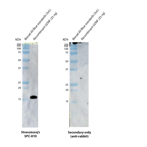
Western blot analysis with Stressmarq’s anti-GDNF polyclonal antibody (SPC-810) showing detection of human recombinant GDNF protein (SinoBiological 10561-HNAE). This recombinant protein has a predicted molecular weight of 15.2 kDa. Block: 5% skim milk. Primary Antibody: Rabbit Anti-GDNF Polyclonal Antibody (SPC-810) at 1:5000 for 2 hours at RT. Secondary Antibody: Goat anti-rabbit IgG:HRP at 1:5000 for 1 hour at RT. Color Development: Chemiluminescent for HRP (Moss) for 3 min in RT. Exposed 9.122 seconds.

Western blot analysis with Stressmarq’s anti-GDNF polyclonal antibody (SPC-810) showing detection of GDNF protein in rat brain lysate (prepared in-house). Rat GNDF protein with signal and pro-peptide segments has a predicted molecular weight of 23.6 kDa (UniProt Q07731). Block: 5% skim milk. Primary Antibody: Rabbit Anti-GDNF Polyclonal Antibody (SPC-810) at 1:1000 for 2 hours at RT. Secondary Antibody: Goat anti-rabbit IgG:HRP at 1:5000 for 1 hour at RT. Color Development: Chemiluminescent for HRP (Moss) for 3 min in RT. Exposed 7.299 seconds. NOTE secondary antibody shows some non-specific binding in the control, but not to the GNDF protein.


Reviews
There are no reviews yet.