Properties
| Storage Buffer | PBS pH7.4, 50% glycerol, 0.09% sodium azide *Storage buffer may change when conjugated |
| Storage Temperature | -20ºC, Conjugated antibodies should be stored according to the product label |
| Shipping Temperature | Blue Ice or 4ºC |
| Purification | Protein G Purified |
| Clonality | Monoclonal |
| Clone Number | S74 |
| Isotype | IgG1 |
| Specificity | Detects ~220kDa in extracts of cells transiently transfected with TrpM7. No cross-reactivity against TrpM6. |
| Cite This Product | StressMarq Biosciences Cat# SMC-316, RRID: AB_2208751 |
| Certificate of Analysis | 1 µg/ml of SMC-316 was sufficient for detection of TrpM7 in 10 µg of COS cell lysate transiently transfected with TprM7 by colorimetric immunoblot analysis using Goat anti-mouse IgG:HRP as the secondary antibody. |
Biological Description
| Alternative Names | CHAK antibody, CHAK1 antibody, Channel kinase 1 antibody, Channel-kinase 1 antibody, Long transient receptor potential channel 7 antibody, LTrpC-7 antibody, LTRPC7 antibody, Transient receptor potential cation channel subfamily M member 7 antibody, TRP PLIK antibody, TRPM7 antibody, TRPM7_HUMAN antibody |
| Research Areas | ALS Disease, Alzheimer's Disease, Ion Channels, Neurodegeneration, Neuroscience, Parkinson's Disease, Transient Receptor Potential (TRP) Channels |
| Cellular Localization | Membrane |
| Accession Number | NP_001157797.1 |
| Gene ID | 58800 |
| Swiss Prot | Q923J1 |
| Scientific Background | TRPs, mammalian homologs of the Drosophila transient receptor potential (trp) protein, are ion channels that are thought to mediate capacitative calcium entry into the cell. TRP-PLIK is a protein that is both an ion channel and a kinase. As a channel, it conducts calcium and monovalent cations to depolarize cells and increase intracellular calcium. As a kinase, it is capable of phosphorylating itself and other substrates. The kinase activity is necessary for channel function, as shown by its dependence on intracellular ATP and by the kinase mutants (1, 2). |
| References |
1. Brauchi S., Krapivinsky G., Krapivinsky L., Clapham D.E. (2008) Proc Natl Acad Sci USA. 105(24): 8304-8308. 2. Numata T, Okada Y. (2008) J Biol Chem. 283(22): 15097-15103. |
Product Images
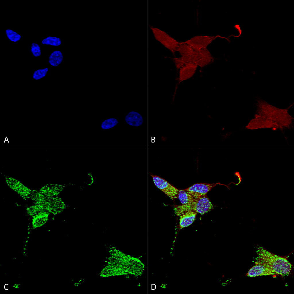
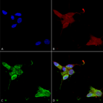
Immunocytochemistry/Immunofluorescence analysis using Mouse Anti-TrpM7 Monoclonal Antibody, Clone S74 (SMC-316). Tissue: Neuroblastoma cells (SH-SY5Y). Species: Human. Fixation: 4% PFA for 15 min. Primary Antibody: Mouse Anti-TrpM7 Monoclonal Antibody (SMC-316) at 1:50 for overnight at 4°C with slow rocking. Secondary Antibody: AlexaFluor 488 at 1:1000 for 1 hour at RT. Counterstain: Phalloidin-iFluor 647 (red) F-Actin stain; Hoechst (blue) nuclear stain at 1:800, 1.6mM for 20 min at RT. (A) Hoechst (blue) nuclear stain. (B) Phalloidin-iFluor 647 (red) F-Actin stain. (C) TrpM7 Antibody (D) Composite.
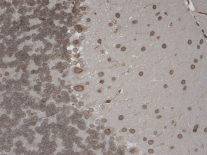
Immunohistochemistry analysis using Mouse Anti-TrpM7 Monoclonal Antibody, Clone S74 (SMC-316). Tissue: Brain Slice. Species: Mouse. Fixation: 10% Formalin Solution for 12-24 hours at RT. Primary Antibody: Mouse Anti-TrpM7 Monoclonal Antibody (SMC-316) at 1:1000 for 1 hour at RT. Secondary Antibody: HRP/DAB Detection System: Biotinylated Goat Anti-Mouse, Streptavidin Peroxidase, DAB Chromogen (brown) for 30 minutes at RT. Counterstain: Mayer Hematoxylin (purple/blue) nuclear stain at 250-500 µl for 5 minutes at RT. Localization: Nuclear staining of both neurons and glia.
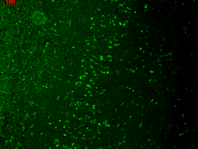
Immunohistochemistry analysis using Mouse Anti-TrpM7 Monoclonal Antibody, Clone S74 (SMC-316). Tissue: hippocampus. Species: Human. Fixation: Bouin’s Fixative and paraffin-embedded. Primary Antibody: Mouse Anti-TrpM7 Monoclonal Antibody (SMC-316) at 1:1000 for 1 hour at RT. Secondary Antibody: FITC Goat Anti-Mouse (green) at 1:50 for 1 hour at RT.
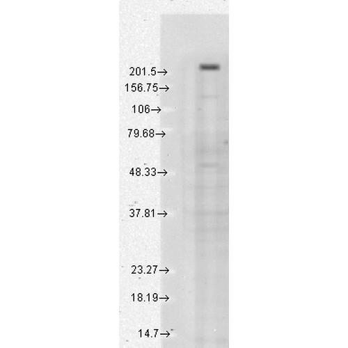
Western Blot analysis of Human cell line mix (A431, A549, HCT116, HeLa, HEK293, HepG2, HL-60, HUVEC, Jurkat, MCF7, PC3, T98G) showing detection of TrpM7 protein using Mouse Anti-TrpM7 Monoclonal Antibody, Clone S74 (SMC-316). Load: 15 µg. Block: 1.5% BSA for 30 minutes at RT. Primary Antibody: Mouse Anti-TrpM7 Monoclonal Antibody (SMC-316) at 1:1000 for 2 hours at RT. Secondary Antibody: Sheep Anti-Mouse IgG: HRP for 1 hour at RT.



StressMarq Biosciences :
Based on validation through cited publications.