| Product Name | VDAC1 Antibody |
| Description |
Mouse Anti-Human VDAC1 Monoclonal IgG2a |
| Species Reactivity | Human, Mouse, Rat |
| Applications | WB, IHC, ICC/IF |
| Antibody Dilution | WB (1:1000); optimal dilutions for assays should be determined by the user. |
| Host Species | Mouse |
| Immunogen Species | Human |
| Immunogen | Fusion protein amino acids 1-283 (full-length) of human VDAC1. Mouse: 98% identity (279/283 amino acids identical). Rat: 98% identity (279/283 amino acids identical) >60% identity with VDAC2 and VDAC3. |
| Concentration | 1 mg/ml |
| Conjugates |
APC, ATTO 390, ATTO 488, ATTO 594, Biotin, FITC, HRP, PerCP, RPE, Unconjugated |
| Field of Use | Not for use in humans. Not for use in diagnostics or therapeutics. For in vitro research use only. |
Properties
| Storage Buffer | PBS pH 7.4, 50% glycerol, 0.1% sodium azide *Storage buffer may change when conjugated |
| Storage Temperature | -20ºC, Conjugated antibodies should be stored according to the product label |
| Shipping Temperature | Blue Ice or 4ºC |
| Purification | Protein G Purified |
| Clonality | Monoclonal |
| Clone Number | N152B/23 (Formerly sold as S152B-23) |
| Isotype | IgG2a |
| Specificity | Detects ~30kDa. Does not cross-react with VDAC2 or VDAC3 (based on KO validation results). |
| Cite This Product | StressMarq Biosciences Cat# SMC-456, RRID: AB_2701955 |
| Certificate of Analysis | 1 µg/ml of SMC-456 was sufficient for detection of VDAC1 in 20 µg of rat brain lysate by colorimetric immunoblot analysis using Goat anti-mouse IgG:HRP as the secondary antibody. |
Biological Description
| Alternative Names | Voltage Dependent Anion Channel 1 Antibody, Porin Antibody, Voltage dependent anion selective channel protein 1 Antibody, Voltage-dependent anion-selective channel protein 1 Antibody, hVDAC1 Antibody, MGC111064 Antibody, Mitochondrial Porin Antibody, Outer mitochondrial membrane protein porin 1 Antibody, Plasmalemmal porin Antibody, Porin 31HL Antibody, Porin 31HM Antibody, PORIN-31-HL Antibody, VDAC 1 Antibody, VDAC Antibody, VDAC-1 Antibody |
| Research Areas | Cancer, Cell Signaling, Ion Channels, Neuroscience |
| Cellular Localization | Cell membrane, Mitochondrion, Mitochondrion outer membrane |
| Accession Number | NP_003365.1. |
| Gene ID | 7416 |
| Swiss Prot | P21796 |
| Scientific Background | Voltage-dependent anion-selective channel protein 1 (also known as VDAC, VDAC1 or outer mitochondrial membrane protein porin 1) is the the outer mitochondrial membrane receptor for hexokinase and BCL2L1. VDAC forms a channel through the mitochondrial membrane and is involved in small molecule diffusion, cell volume regulation and apoptosis. VDAC may participate in the formation of the permeability transition pore complex (PTPC), which is responsible for the release of mitochondrial products that triggers apoptosis. |
Product Images

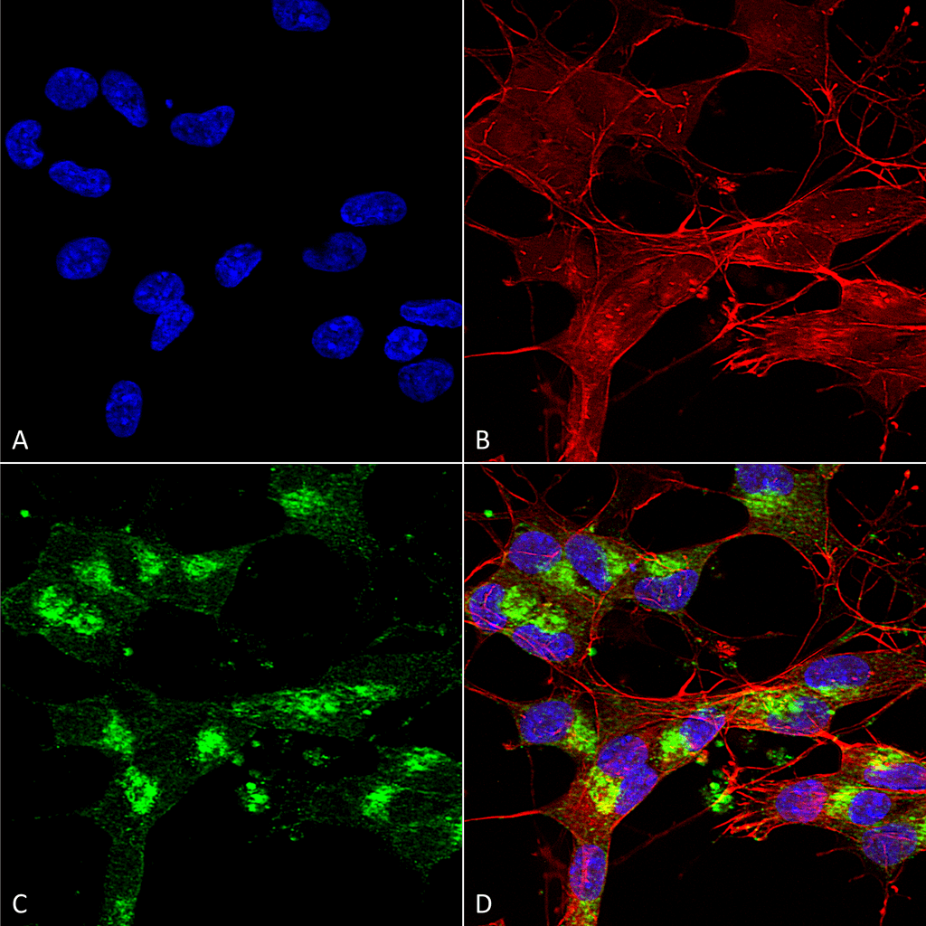
Immunocytochemistry/Immunofluorescence analysis using Mouse Anti-VDAC1 Monoclonal Antibody, Clone N152B/23 (SMC-456). Tissue: Neuroblastoma cells (SH-SY5Y). Species: Human. Fixation: 4% PFA for 15 min. Primary Antibody: Mouse Anti-VDAC1 Monoclonal Antibody (SMC-456) at 1:100 for overnight at 4°C with slow rocking. Secondary Antibody: AlexaFluor 488 at 1:1000 for 1 hour at RT. Counterstain: Phalloidin-iFluor 647 (red) F-Actin stain; Hoechst (blue) nuclear stain at 1:800, 1.6mM for 20 min at RT. (A) Hoechst (blue) nuclear stain. (B) Phalloidin-iFluor 647 (red) F-Actin stain. (C) VDAC1 Antibody (D) Composite.

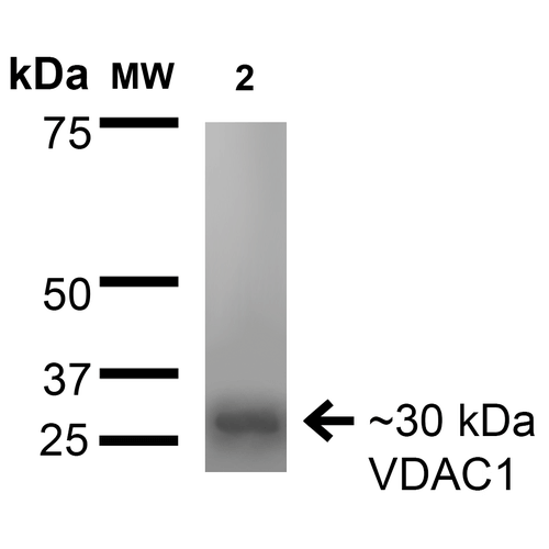
Western Blot analysis of Rat Brain Membrane showing detection of ~30 kDa VDAC1 protein using Mouse Anti-VDAC1 Monoclonal Antibody, Clone N152B/23 (SMC-456). Lane 1: Molecular Weight Ladder. Lane 2: Rat Brain Membrane. Load: 15 µg. Block: 2% BSA and 2% Skim Milk in 1X TBST. Primary Antibody: Mouse Anti-VDAC1 Monoclonal Antibody (SMC-456) at 1:200 for 16 hours at 4°C. Secondary Antibody: Goat Anti-Mouse IgG: HRP at 1:1000 for 1 hour RT. Color Development: ECL solution for 6 min in RT. Predicted/Observed Size: ~30 kDa.

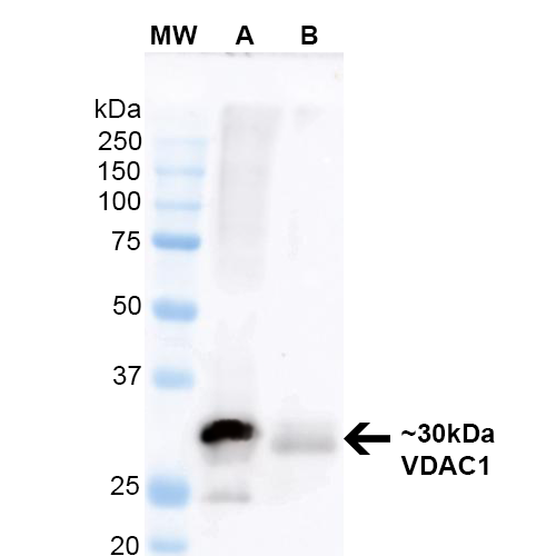
Western Blot analysis of Mouse Brain and Human RT-4 lysates showing detection of ~30 kDa VDAC1 protein using Mouse Anti-VDAC1 Monoclonal Antibody, Clone N152B/23 (SMC-456). Lane 1: Molecular Weight Ladder. Lane A: Mouse Brain Lysate. Lane B: Human RT-4 Lysate. Block: 5% Skim milkin TBST . Primary Antibody: Mouse Anti-VDAC1 Monoclonal Antibody (SMC-456) at 1:200 for 60 min at RT. Secondary Antibody: Goat Anti-Mouse IgG: HRP at 1:2000 for 60 min at RT. Color Development: ECL solution for 5 min in RT. Predicted/Observed Size: ~30 kDa.


Stephani Davis :
Works great for westerns. The first time I tried it I used 1:1000 and it burned into the membrane (10ug mouse brain lysate). 1:10,000 worked well.