| Product Name | 20S Proteasome Activity Kit |
| Description |
Fluorometric detection of purified 20S proteasome activity |
| Species Reactivity | Species Independent |
| Platform | Microplate |
| Sample Types | Purified Proteins |
| Detection Method | Fluorometric Assay |
| Assay Type | Continuous Kinetic Assay |
| Utility | Activity kit used to screen potential proteasome inhibitors and activators, and to measure and quantify 20S proteasome activity in continuous kinetic and end-point assays. |
| Incubation Time | 15-30 minutes |
| Number of Samples | 40 samples in duplicate using the plate format, or 100x 50µL micro-cuvette based assays |
| Other Resources | Kit Booklet , MSDS |
| Field of Use | Not for use in humans. Not for use in diagnostics or therapeutics. For in vitro research use only. |
Properties
| Storage Temperature | 4ºC | |||||||||||||||||||||
| Shipping Temperature | Blue Ice | |||||||||||||||||||||
| Product Type | Activity Kits | |||||||||||||||||||||
| Assay Overview | The 20S Proteasome Activity kit facilitates the rapid, robust measurement of proteasome activity in vitro. The kit utilises high purity, fluorogenic substrate Suc-LLVY-AMC together with suitable calibration standards and controls for the accurate and sensitive assessment of proteasome chymotrypsin-like (β5) activity. Continuous kinetic or end-point assays can be performed in micro-cuvettes or in 96-well plate format for multi-sample analysis. Contains sufficient materials for one full 96-well plate assay or up to 100x 50µL micro-cuvette based assays to be run. | |||||||||||||||||||||
| Kit Overview |
|
|||||||||||||||||||||
| Cite This Product | 20S Proteasome Activity Kit (StressMarq Biosciences Inc., Victoria BC CANADA, Catalog # SKT-133) |
Biological Description
| Alternative Names | Chymotrypsin-like (β5) Activity Kit |
| Research Areas | Apoptosis, Cancer, Cardiovascular System, Cell Signaling, Neurodegeneration, Neuroscience, Post-translational Modifications |
| Scientific Background | Proteasomes are non-lysosomal proteolytic complexes localised primarily in the cytoplasm and in the nucleus of eukaryotic cells. They are responsible for the ubiquitin-mediated degradation of short half-life proteins and peptides that are involved in essential cellular processes including cell-cycle regulation, apoptosis and transcriptional regulation, innate immunity and antigen processing, and in the removal of redundant or damaged proteins. As such protein degradation by the ubiquitin-proteasome pathway has a major regulatory function for proliferation activity and survival of both normal and malignant cells, and its dysfunction has been implicated in a wide range of other disease processes including neurodegenerative, cardiovascular and metabolic disorders. The 26S proteasome structure is composed of a 20S proteasome catalytic core complex and one or two 19S (PA700) regulatory subcomplexes. The 20S core comprises two copies of 14 subunits (7 alpha subunits and 7 beta subunits) arranged in a α7β7β7α7 cylindrical array. Proteolytic activities are determined by the β1 (caspase-like), β2 (trypsin-like) and β5 (chymotrypsin-like) subunits, access to which is guarded by the α-subunits. The 19S regulatory unit consists of six ATPase and at least ten non-ATPase subunits that are required for ubiquitinated protein binding, deubiquitination, substrate unfolding and translocation to the 20S catalytic core. Varying catalytic subunit composition (β1, β1i; β2, β2i; β5, β5i) results in a variety of possible subtypes from full constitutive proteasome (β1, β2, β5) through mixed populations to full inducible / immunoproteasome (β1i, β2i, β5i). Alternative regulatory complexes such as the PA200 and 11S proteasome activators confer different substrate specificities and activity compared to the 19S regulator. |
| References |
1. Saeki, Y. & Tanaka, K. Methods in molecular biology (Clifton, N.J.) 832, 315–37 (2012). 2. Frankland-Searby, S. & Bhaumik, S. R. Biochimica et biophysica acta 1825, 64–76 (2012). 3. Basler, M., Kirk, C. J. & Groettrup, M. Current opinion in immunology 1–7 (2012). 4. Löw, P. General and comparative endocrinology 172, 39–43 (2011). 5. Dennissen, F. J. a, Kholod, N. & Van Leeuwen, F. W. Progress in neurobiology 96, 190–207 (2012). 6. Geng, F., Wenzel, S. & Tansey, W. P. Annual review of biochemistry 81, 177–201 (2012). |
Product Images

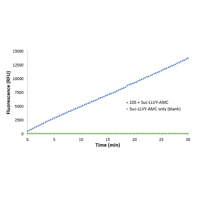
Continuous kinetic enzyme activity assay showing the time course of control 20S proteasome activity with Suc-LLVY-AMC substrate using the StressXpress® 20S Proteasome Activity Kit. 20S proteasome (~2.5nM) was incubated with 100µM Suc-LLVY-AMC in proteasome assay buffer for 30 minutes at room temperature alongside a substrate only (blank) control reaction. Fluorescence measurements (RFU) were taken at 30 second intervals and plotted vs. time.


Reviews
There are no reviews yet.