Alpha Synuclein Protein

Mouse Recombinant Alpha Synuclein Pre-formed Fibrils (Type 1)

Catalog No. SPR-324

5 out of 5 based on 1 customer rating
Expression System E. coli
Tags No tag
SKU: SPR-324 Category:

Clear
SPR-324_Alpha-Synuclein-Protein-Preformed-Fibrils-ICC-IF-1.png
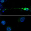
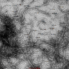
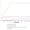
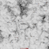
IHC of mouse brain injected with alpha synuclein monomers and fibrilsIHC of mouse brain injected with alpha synuclein monomers and fibrilsIHC of Sprague-Dawley rat brain injected with Type 1 mouse alpha synuclein Pre-formed Fibrils (SPR-324)Mouse hippocampal neurons treated with mouse alpha synuclein PFFs show phosphorylationTEM of Type 1 mouse alpha synuclein Pre-formed Fibrils (SPR-324).Mouse Type 1 alpha synuclein Pre-formed Fibrils (SPR-324) seed the formation of new alpha Synuclein fibrils from the pool of alpha synuclein monomers (SPR-323) as shown by increased fluorescence intensity by Thioflavin T over time.TEM of Type 1 mouse alpha synuclein Pre-formed Fibrils (SPR-324).SDS-Page of Type 1 Mouse Recombinant Alpha Synuclein Protein Pre-formed Fibrils (SPR-324)
Product Name Alpha Synuclein Protein
Description

Mouse Recombinant Alpha Synuclein Pre-formed Fibrils (Type 1)

Applications WB, SDS-PAGE, In vivo assay, In vitro assay
Concentration 2 mg/ml or 5 mg/ml
Conjugates No tag
Dylight 488
Overview:

  • High fluorescence yield
  • High photostability
  • Less pH-sensitive
  • Excellent batch-to-batch reproducibility
  • Stringently QC tested
  • Molecular weight: 1011 g/mol

Dylight 488 Datasheet

Dylight 488 Fluorophore Absorption and Emission Spectrum Optical Properties:

λex = 493 nm

λem = 518 nm

εmax = 7.0×10⁴

Laser = 488 nm


APC/Cy7
Overview:

  • High quantum yield
  • Excellent batch-to-batch reproducibility
  • Stringently QC tested

APC-Cy7 Datasheet


ACP-Cy7 Fluorophore Absorption and Emission Spectrum Optical Properties:

λex = 652 nm

λem = 790 nm

Laser = 594 or 633 nm


  Dylight 350
Overview:

  • High fluorescence intensity
  • High photostability
  • Less pH-sensitive
  • Excellent solubility in water
  • Stringently QC tested
  • Excellent batch-to-batch reproducibility
  • Molecular weight: 874 g/mol

Dylight 350 Datasheet

Dylight 350 Fluorophore Absorption and Emission Spectrum Optical Properties:

λex = 353 nm

λem = 432 nm

εmax = 1.5×10⁴


  Dylight 405
Overview:

  • High fluorescence intensity
  • High photostability
  • Less pH-sensitive
  • Excellent batch-to-batch reproducibility
  • Stringently QC tested
  • Molecular weight: 793 g/mol

Dylight 405 Datasheet

Dylight 405 Fluorophore Absorption and Emission Spectrum Optical Properties:

λex = 400 nm

λem = 420 nm

εmax = 3.0×10⁴

Laser = 405 nm


Dylight 594
Overview:

  • High fluorescence yield
  • High photostability
  • Less pH-sensitive
  • Excellent batch-to-batch reproducibility
  • Stringently QC tested
  • Molecular weight: 1078 g/mol

Dylight 594 Datasheet

Dylight 594 Fluorophore Absorption and Emission Spectrum Optical Properties:

λex = 593 nm

λem = 618 nm

εmax = 8.0×10⁴

Laser = 526 nm


 Dylight 633
Overview:

  • High fluorescence yield
  • High photostability
  • Less pH-sensitive
  • Excellent batch-to-batch reproducibility
  • Stringently QC tested
  • Molecular weight: 1066 g/mol

Dylight 633 Datasheet

Dylight 633 Fluorophore Absorption and Emission Spectrum Optical Properties:

λex = 638 nm

λem = 658 nm

εmax = 1.7×10⁵

Laser = 633 nm


 PerCP 
Overview:

  • Peridinin-Chlorophyll-Protein Complex
  • Small phycobiliprotein
  • Isolated from red algae
  • Large stokes shift (195 nm)
  • Molecular Weight: 35 kDa

PerCP Datasheet

 PerCP Fluorophore Absorption and Emission Spectrum Optical Properties:

λex = 482 nm

λem = 677 nm

εmax = 1.96 x 10⁶

Laser = 488 nm


 PE/ATTO 594
PE/ATTO 594 is a tandem conjugate, where PE is excited at 535 nm and transfers energy to ATTO 594 via FRET (fluorescence resonance energy transfer), which emits at 627 nm.
Overview:

  • High fluorescence yield
  • High photostability
  • Very hydrophilic
  • Excellent solubility in water
  • Very little aggregation

PE/ATTO 594 Datasheet

PE-ATTO 594 Fluorophore Conjugate Excitation and Emission Spectra Optical Properties:

λex = 535 nm

λem = 627 nm

Laser = 488 to 561 nm


  FITC (Fluorescein)
Overview:

  • Excellent fluorescence quantum yield
  • High rate of photobleaching
  • Good solubility in water
  • Broad emission spectrum
  • pH dependent spectra
  • Molecular formula: C20H12O5
  • Molar mass: 332.3 g/mol

FITC-Fluorescent-conjugate

FITC Fluorescein Fluorophore Excitation and Emission Spectra Optical Properties:

λex = 494 nm

λem = 520 nm

εmax = 7.3×10⁴

Φf = 0.92

τfl = 5.0 ns

Brightness = 67.2

Laser = 488 nm

Filter set = FITC


 ATTO 700
Overview:

  • High fluorescence yield
  • Excellent thermal and photostability
  • Quenched by electron donors
  • Very hydrophilic
  • Good solubility in polar solvents
  • Zwitterionic dye
  • Molar Mass: 575 g/mol

ATTO 700 Datasheet

 ATTO 700 Fluorophore Absorption and Emission Spectrum Optical Properties:

λex = 700 nm

λem = 719 nm

εmax = 1.25×10⁵

Φf = 0.25

τfl = 1.6 ns

Brightness = 31.3

Laser = 676 nm

Filter set = Cy®5.5


 ATTO 680
Overview:

  • High fluorescence yield
  • Excellent thermal and photostability
  • Quenched by electron donors
  • Very hydrophilic
  • Good solubility in polar solvents
  • Zwitterionic dye
  • Molar Mass: 631 g/mol

ATTO 680 Datasheet

 ATTO 680 Fluorophore Absorption and Emission Spectrum Optical Properties:

λex = 680 nm

λem = 700 nm

εmax = 1.25×10⁵

Φf = 0.30

τfl = 1.7 ns

Brightness = 37.5

Laser = 633 – 676 nm

Filter set = Cy®5.5


 ATTO 655
Overview:

  • High fluorescence yield
  • High thermal and photostability
  • Excellent ozone resistance
  • Quenched by electron donors
  • Very hydrophilic
  • Good solubility in polar solvents
  • Zwitterionic dye
  • Molar Mass: 634 g/mol

ATTO 655 Datasheet

ATTO 655 Fluorophore Absorption and Emission Spectrum Optical Properties:

λex = 663 nm

λem = 684 nm

εmax = 1.25×10⁵

Φf = 0.30

τfl = 1.8 ns

Brightness = 37.5

Laser = 633 – 647 nm

Filter set = Cy®5


 ATTO 633
Overview:

  • High fluorescence yield
  • High thermal and photostability
  • Moderately hydrophilic
  • Good solubility in polar solvents
  • Stable at pH 4 – 11
  • Cationic dye, perchlorate salt
  • Molar Mass: 652.2 g/mol

ATTO 633 Datasheet

ATTO 633 Fluorophore Absorption and Emission Spectrum Optical Properties:

λex = 629 nm

λem = 657 nm

εmax = 1.3×10⁵

Φf = 0.64

τfl = 3.2 ns

Brightness = 83.2

Laser = 633 nm

Filter set = Cy®5


 ATTO 594
Overview:

  • High fluorescence yield
  • High photostability
  • Very hydrophilic
  • Excellent solubility in water
  • Very little aggregation
  • New dye with net charge of -1
  • Molar Mass: 1137 g/mol

ATTO 594 Datasheet

 ATTO 594 Fluorophore Excitation and Emission Spectrum Optical Properties:

λex = 601 nm

λem = 627 nm

εmax = 1.2×10⁵

Φf = 0.85

τfl = 3.5 ns

Brightness = 102

Laser = 594 nm

Filter set = Texas Red®


 ATTO 565
Overview:

  • High fluorescence yield
  • High thermal and photostability
  • Good solubility in polar solvents
  • Excellent solubility in water
  • Very little aggregation
  • Rhodamine dye derivative
  • Molar Mass: 611 g/mol

ATTO 565 Datasheet

 ATTO 565 Fluorophore Excitation and Emission Spectra Optical Properties:

λex = 563 nm

λem = 592 nm

εmax = 1.2×10⁵

Φf = 0.9

τfl = 3.4 n

Brightness = 10

Laser = 532 nm

Filter set = TRITC


  ATTO 488
Overview:

  • High fluorescence yield
  • High photostability
  • Very hydrophilic
  • Excellent solubility in water
  • Very little aggregation
  • New dye with net charge of -1
  • Molar Mass: 804 g/mol 

ATTO 488 Datasheet

  ATTO 488 Fluorophore Excitation and Emission Spectra Optical Properties:

λex = 501 nm

λem = 523 nm

εmax = 9.0×10⁴

Φf = 0.80

τfl = 4.1 ns

Brightness = 72

Laser = 488 nm

Filter set = FITC


  ATTO 390
Overview:

  • High fluorescence yield
  • Large Stokes-shift (89 nm)
  • Good photostability
  • Moderately hydrophilic
  • Good solubility in polar solvents
  • Coumarin derivate, uncharged
  • Low molar mass: 343.42 g/mol 

ATTO 390 Datasheet

ATTO 390 Fluorescent Dye Excitation and Emission Spectra Optical Properties:

λex = 390 nm

λem = 479 nm

εmax = 2.4×10⁴

Φf = 0.90

τfl = 5.0 ns

Brightness = 21.6

Laser = 365 or 405 nm


APC (Allophycocyanin)
Overview:

  • High quantum yield
  • Large phycobiliprotein
  • 6 chromophores per molecule
  • Isolated from red algae
  • Molecular Weight: 105 kDa

APC Datasheet

 APC Fluorophore Absorption and Emission Spectrum Optical Properties:

λex = 650 nm

λem = 660 nm

εmax = 7.0×10⁵

Φf = 0.68

Brightness = 476

Laser = 594 or 633 nm

Filter set = Cy®5


Streptavidin

Properties:

  • Homo-tetrameric protein purified from Streptomyces avidinii which binds four biotin molecules with extremely high affinity
  • Molecular weight: 53 kDa
  • Formula: C10H16N2O3S
  • Applications: Western blot, immunohistochemistry, and ELISA

Streptavidin Datasheet

BiotinBiotin Conjugate Structure

Properties:

  • Binds tetrameric avidin proteins including Streptavidin and neuravidin with very high affinity
  • Molar mass: 244.31 g/mol
  • Formula: C10H16N2O3S
  • Applications: Western blot, immunohistochemistry, and ELISA

Biotin Datasheet

HRP (Horseradish peroxidase)

Properties:

  • Enzymatic activity is used to amplify weak signals and increase visibility of a target
  • Readily combines with hydrogen peroxide (H2O2) to form HRP-H2O2 complex which can oxidize various hydrogen donors
  • Catalyzes the conversion of:
    • Chromogenic substrates (e.g. TMB, DAB, ABTS) into coloured products
    • Chemiluminescent substrates (e.g. luminol and isoluminol) into light emitting products via enhanced chemiluminescence (ECL)
    • Fluorogenic substrates (e.g. tyramine, homovanillic acid, and 4-hydroxyphenyl acetic acid) into fluorescent products
  • High turnover rate enables rapid generation of a strong signal
  • 44 kDa glycoprotein
  • Extinction coefficient: 100 (403 nm)
  • Applications: Western blot, immunohistochemistry, and ELISA

HRP Datasheet

AP (Alkaline Phosphatase)

Properties:

  • Broad enzymatic activity for phosphate esters of alcohols, amines, pyrophosphate, and phenols
  • Commonly used to dephosphorylate the 5’-termini of DNA and RNA to prevent self-ligation
  • Catalyzes the conversion of:
    • Chromogenic substrates (e.g. pNPP, naphthol AS-TR phosphate, BCIP) into coloured products
    • Fluorogenic substrates (e.g. 4-methylumbelliferyl phosphate) into fluorescent products
  • Molecular weight: 140 kDa
  • Applications: Western blot, immunohistochemistry, and ELISA

AP Datasheet

  R-PE (R-Phycoerythrin)
Overview:

  • Broad excitation spectrum
  • High quantum yield
  • Photostable
  • Member of the phycobiliprotein family
  • Isolated from red algae
  • Excellent solubility in water
  • Molecular Weight: 250 kDa

R-PE Datasheet

 R-PE Fluorophore Excitation and Emission Spectra Optical Properties:

λex = 565 nm

λem = 575 nm

εmax = 2.0×10⁶

Φf = 0.84

Brightness = 1.68 x 10³

Laser = 488 to 561 nm

Filter set = TRITC


Nature Recombinant
Species Mouse
Expression System E. coli
Amino Acid Sequence MDVFMKGLSK AKEGVVAAAE KTKQGVAEAA GKTKEGVLYV GSKTKEGVVH GVTTVAEKTK EQVTNVGGAV VTGVTAVAQK TVEGAGNIAA ATGFVKKDQM GKGEEGYPQE GILEDMPVDP GSEAYEMPSE EGYQDYEPEA
Purity >95%
Other Resources Sonication Protocol
Protein Length Full Length
Protein Size ~14.46 kDa
Biological Activity Endogenous alpha-synuclein phosphorylation. 100 µM alpha synuclein protein monomer (SPR-323) seeded with 10 uM alpha synuclein protein PFF (SPR-324) in 25 µM Thioflavin T (PBS pH 7.4, 100 µl reaction volume) generated an increased fluorescence intensity after incubation at 37°C with shaking at 600 rpm. Fluorescence was measured by excitation at 450 nm and emission at 485 nm on a Molecular Devices Gemini XPS microplate reader.
Field of Use Not for use in humans. Not for use in diagnostics or therapeutics. For in vitro research use only.

Properties

Storage Buffer PBS pH 7.4
Storage Temperature -80ºC
Shipping Temperature Dry Ice. Shipping note: Product will be shipped separately from other products purchased in the same order.
Purification Ion-exchange Purified
Cite This Product Mouse Recombinant Alpha Synuclein Protein (StressMarq Biosciences Inc., Victoria BC CANADA, Catalog # SPR-324)
Certificate of Analysis Certified >95% pure using SDS-PAGE analysis. Low endotoxin <5 EU/mL @ 2mg/mL.
Other Relevant Information For best results, sonicate immediately prior to use. Refer to the Neurodegenerative Protein Handling Instructions on our website, or the product datasheet for further information. Monomer source is catalog# SPR-323.

Biological Description

Alternative Names Alpha synuclein PFFs, Alpha synuclein PFF, Alpha synuclein aggregates, Alpha synuclein protein aggregates, Alpha synuclein aggregates, Alpha-synuclein protein, Non-A beta component of AD amyloid protein, Non-A4 component of amyloid precursor protein, NACP protein, SNCA protein, NACP protein, PARK1 protein, SYN protein, Parkinson's disease familial 1 Protein
Research Areas Alzheimer's Disease, Neurodegeneration, Neuroscience, Parkinson's Disease, Synuclein, Tangles & Tau, Multiple System Atrophy
Cellular Localization Cytoplasm, Membrane, Nucleus
Accession Number NP_001035916.1
Gene ID 20617
Swiss Prot O55042
Scientific Background Alpha-Synuclein (SNCA) is expressed predominantly in the brain, where it is concentrated in presynaptic nerve terminals (1). Alpha-synuclein is highly expressed in the mitochondria of the olfactory bulb, hippocampus, striatum and thalamus (2). Functionally, it has been shown to significantly interact with tubulin (3), and may serve as a potential microtubule-associated protein. It has also been found to be essential for normal development of the cognitive functions; inactivation may lead to impaired spatial learning and working memory (4). SNCA fibrillar aggregates represent the major non A-beta component of Alzheimers disease amyloid plaque, and a major component of Lewy body inclusions, and Parkinson's disease. Parkinson's disease (PD) is a common neurodegenerative disorder characterized by the progressive accumulation in selected neurons of protein inclusions containing alpha-synuclein and ubiquitin (5, 6).
References 1. “Genetics Home Reference: SNCA”. US National Library of Medicine. (2013).
2. Zhang L., et al. (2008) Brain Res. 1244: 40-52.
3. Alim M.A., et al. (2002) J Biol Chem. 277(3): 2112-2117.
4. Kokhan V.S., Afanasyeva M.A., Van'kin G. (2012) Behav. Brain. Res. 231(1): 226-230.
5. Spillantini M.G., et al. (1997) Nature. 388(6645): 839-840.
6. Mezey E., et al. (1998) Nat Med. 4(7): 755-757.

Product Images

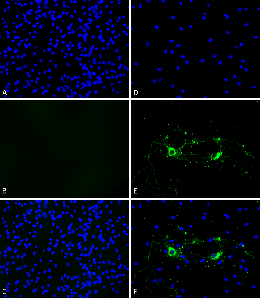
<p>Primary rat hippocampal neurons show lewy body inclusion formation and loss of cells when treated with Type 1 mouse Alpha Synuclein Protein Pre-formed Fibrils (SPR-324) at 4 µg/ml (D-F) on DVI2, but not when treated with a control (A-C). Tissue: Primary hippocampal neurons. Species: Sprague-Dawley rat. Fixation: 3% formaldehyde from PFA for 20 min. Blocker: 1:1 PBS:LiCOR Odyssey Block (LiCOR, 927-40010) and 30 mL/mL of 0.1% triton-X 100 for 30 min. Primary Antibody: Mouse anti-pSer129 Antibody (1:1000) and Rabbit anti-pSer129 (1:800) for 24 hours at 4°C. Secondary Antibody: ATTO 546 Donkey Anti-Mouse (1:700) and ATTO 488 Donkey Anti-Rabbit (1:700) for 1 hour at RT (composite green). Counterstain: Hoechst (blue) nuclear stain at 1:3000 for 1 hour at RT. Localization: Lewy body inclusions. Magnification: 20x.</p>

Primary rat hippocampal neurons show lewy body inclusion formation and loss of cells when treated with Type 1 mouse Alpha Synuclein Protein Pre-formed Fibrils (SPR-324) at 4 µg/ml (D-F) on DVI2, but not when treated with a control (A-C). Tissue: Primary hippocampal neurons. Species: Sprague-Dawley rat. Fixation: 3% formaldehyde from PFA for 20 min. Blocker: 1:1 PBS:LiCOR Odyssey Block (LiCOR, 927-40010) and 30 mL/mL of 0.1% triton-X 100 for 30 min. Primary Antibody: Mouse anti-pSer129 Antibody (1:1000) and Rabbit anti-pSer129 (1:800) for 24 hours at 4°C. Secondary Antibody: ATTO 546 Donkey Anti-Mouse (1:700) and ATTO 488 Donkey Anti-Rabbit (1:700) for 1 hour at RT (composite green). Counterstain: Hoechst (blue) nuclear stain at 1:3000 for 1 hour at RT. Localization: Lewy body inclusions. Magnification: 20x.
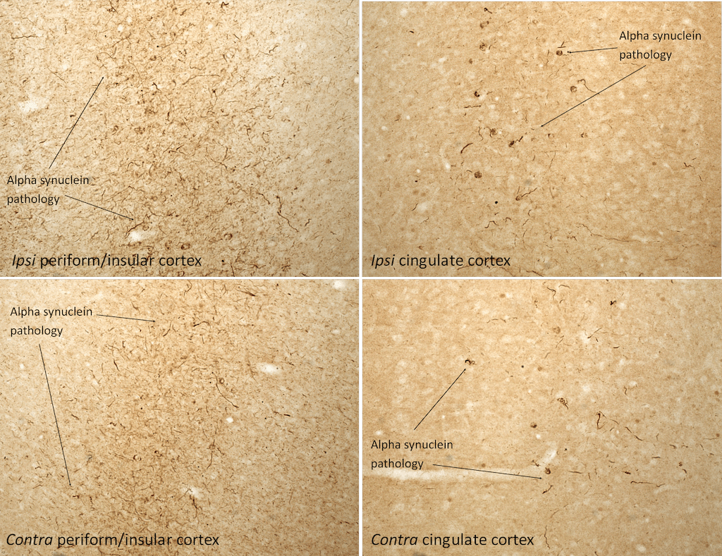
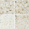
<p>Immunohistochemistry analysis of rat brain injected with Type 1 mouse alpha synuclein PFFs (SPR-324). Species: Female Sprague-Dawley Rat. Rat was injected with 16µg Type 1 mouse alpha synuclein PFFs (SPR-324) in each of 2 injection sites: AP+1.6, ML+2.4, DV-4.2 from skull; and AP-1.4, ML+0.2, DV-2.8 from skull. 30 days post-injection. Fixation: Saline perfusion followed by 4% PFA fixation for 48 hrs. Primary antibody: rabbit monoclonal anti-pSer129 alpha synuclein. Secondary Antibody: Biotin-SP Donkey Anti-Rabbit IgG (H+L) at 1:500 for 2 hours in cold room with shaking. ABC signal amplification, DAB staining. Magnification: 20X. Alpha synuclein pathology is seen in the periform/insular cortex and the cingulate cortex on both the same (ipsi) and opposite (contra) sides as the injection sites.</p>

Immunohistochemistry analysis of rat brain injected with Type 1 mouse alpha synuclein PFFs (SPR-324). Species: Female Sprague-Dawley Rat. Rat was injected with 16µg Type 1 mouse alpha synuclein PFFs (SPR-324) in each of 2 injection sites: AP+1.6, ML+2.4, DV-4.2 from skull; and AP-1.4, ML+0.2, DV-2.8 from skull. 30 days post-injection. Fixation: Saline perfusion followed by 4% PFA fixation for 48 hrs. Primary antibody: rabbit monoclonal anti-pSer129 alpha synuclein. Secondary Antibody: Biotin-SP Donkey Anti-Rabbit IgG (H+L) at 1:500 for 2 hours in cold room with shaking. ABC signal amplification, DAB staining. Magnification: 20X. Alpha synuclein pathology is seen in the periform/insular cortex and the cingulate cortex on both the same (ipsi) and opposite (contra) sides as the injection sites.
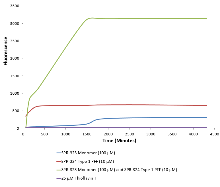
<p>Type 1 alpha synuclein Pre-formed Fibrils (SPR-324) seed the formation of new alpha synuclein fibrils from the pool of alpha synuclein monomers (SPR-323). Thioflavin T is a fluorescent dye that binds to beta sheet-rich structures, such as those in alpha synuclein fibrils. Upon binding, the emission spectrum of the dye experiences a red-shift, and increased fluorescence intensity. Thioflavin T emission curves show increased fluorescence (correlated to alpha synuclein protein aggregation) over time when 10 µM of Type 1 alpha synuclein Pre-formed Fibrils (SPR-324) is combined with 100 µM of alpha synuclein monomer (SPR-323), as compared to Type 1 alpha synuclein Pre-formed Fibrils (SPR-324) or alpha Synuclein monomer (SPR-323) alone. Thioflavin T ex = 450 nm, em = 485 nm.</p>

Type 1 alpha synuclein Pre-formed Fibrils (SPR-324) seed the formation of new alpha synuclein fibrils from the pool of alpha synuclein monomers (SPR-323). Thioflavin T is a fluorescent dye that binds to beta sheet-rich structures, such as those in alpha synuclein fibrils. Upon binding, the emission spectrum of the dye experiences a red-shift, and increased fluorescence intensity. Thioflavin T emission curves show increased fluorescence (correlated to alpha synuclein protein aggregation) over time when 10 µM of Type 1 alpha synuclein Pre-formed Fibrils (SPR-324) is combined with 100 µM of alpha synuclein monomer (SPR-323), as compared to Type 1 alpha synuclein Pre-formed Fibrils (SPR-324) or alpha Synuclein monomer (SPR-323) alone. Thioflavin T ex = 450 nm, em = 485 nm.
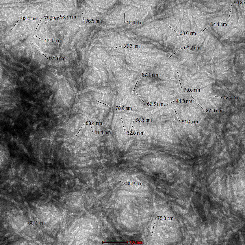
<p>TEM of Type 1 mouse alpha synuclein Pre-formed Fibrils (SPR-324). Image was taken at 100kx magnification.</p>

TEM of Type 1 mouse alpha synuclein Pre-formed Fibrils (SPR-324). Image was taken at 100kx magnification.

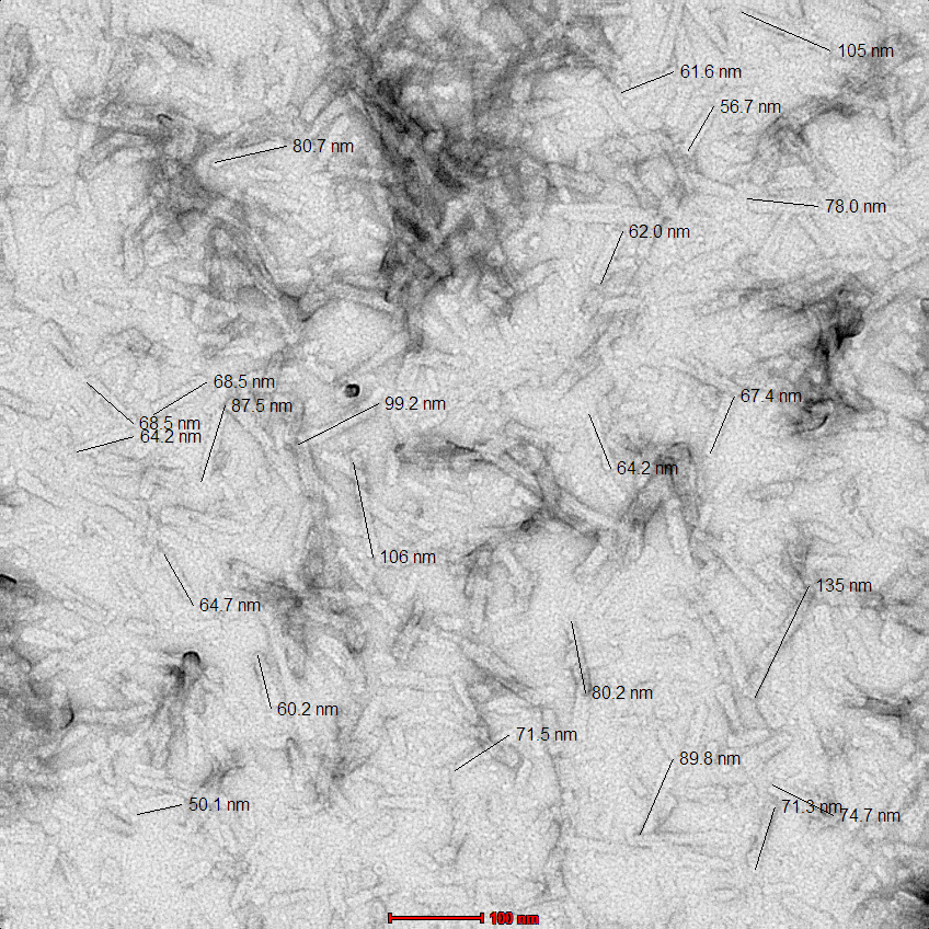
<p>TEM of Type 1 mouse alpha synuclein Pre-formed Fibrils (SPR-324). Fibrils were sonicated and image was taken at 100kx magnification.</p>

TEM of Type 1 mouse alpha synuclein Pre-formed Fibrils (SPR-324). Fibrils were sonicated and image was taken at 100kx magnification.

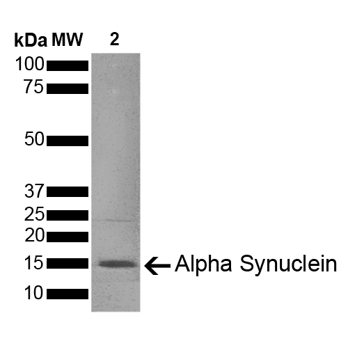
<p>SDS-PAGE of ~14 kDa Type 1 Mouse Recombinant Alpha Synuclein Protein Pre-formed Fibrils (SPR-324). Lane 1: Molecular Weight Ladder (MW). Lane 2: Type 1 Alpha Synuclein Protein Pre-formed Fibrils (2 µg) (SPR-324).</p>

SDS-PAGE of ~14 kDa Type 1 Mouse Recombinant Alpha Synuclein Protein Pre-formed Fibrils (SPR-324). Lane 1: Molecular Weight Ladder (MW). Lane 2: Type 1 Alpha Synuclein Protein Pre-formed Fibrils (2 µg) (SPR-324).

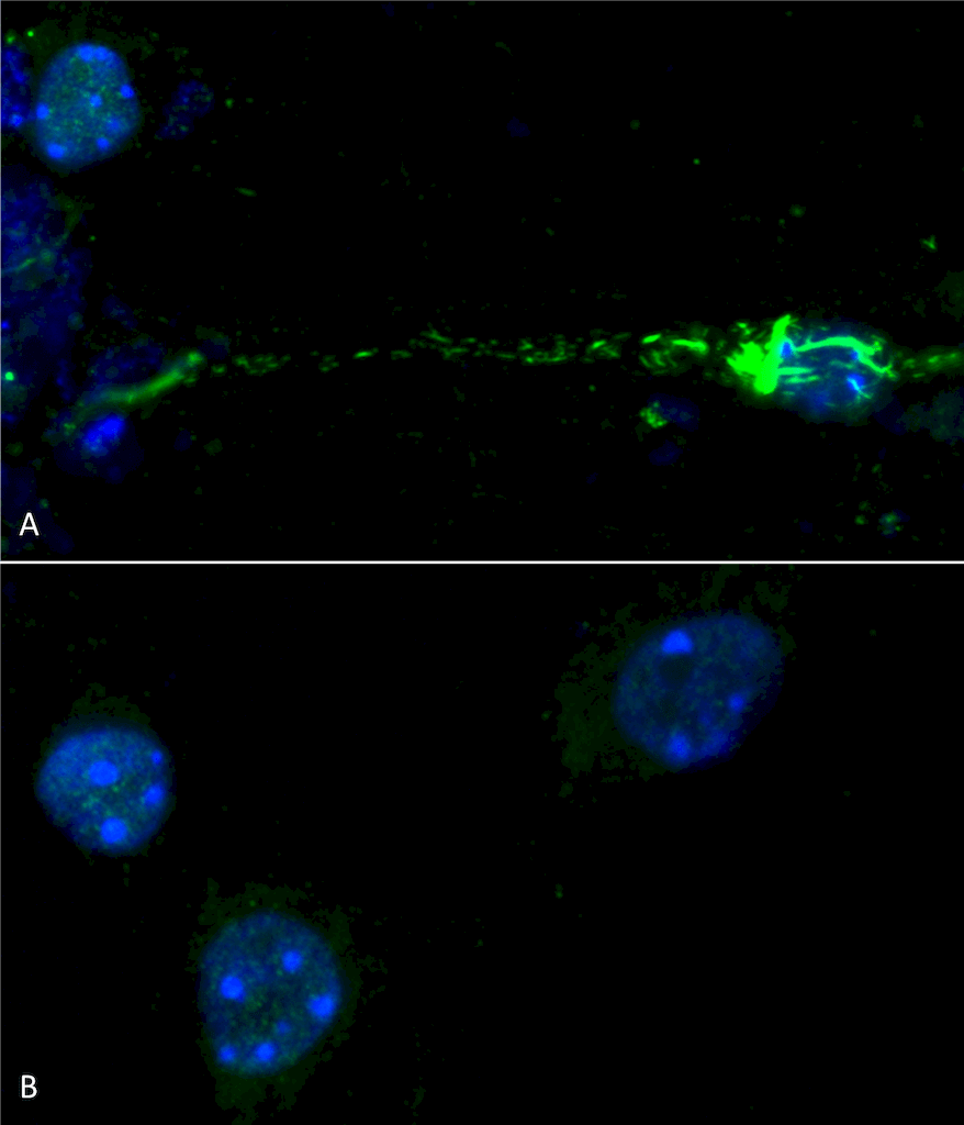
<p>Primary mouse hippocampal neurons treated with 100 nM sonicated mouse alpha synuclein PFFs (SPR-324) (A). Phosphorylated alpha synuclein (detected with pSer129 antibody SPC-742) was visible in perinucleus and neurites compared to untreated control (B). Read the protocol at pabmabs.com/?p=7901. Image courtesy of Trine Rasmussen, Simon Molgaard Jensen at Aarhus University.</p>

Primary mouse hippocampal neurons treated with 100 nM sonicated mouse alpha synuclein PFFs (SPR-324) (A). Phosphorylated alpha synuclein (detected with pSer129 antibody SPC-742) was visible in perinucleus and neurites compared to untreated control (B). Read the protocol at pabmabs.com/?p=7901. Image courtesy of Trine Rasmussen, Simon Molgaard Jensen at Aarhus University.
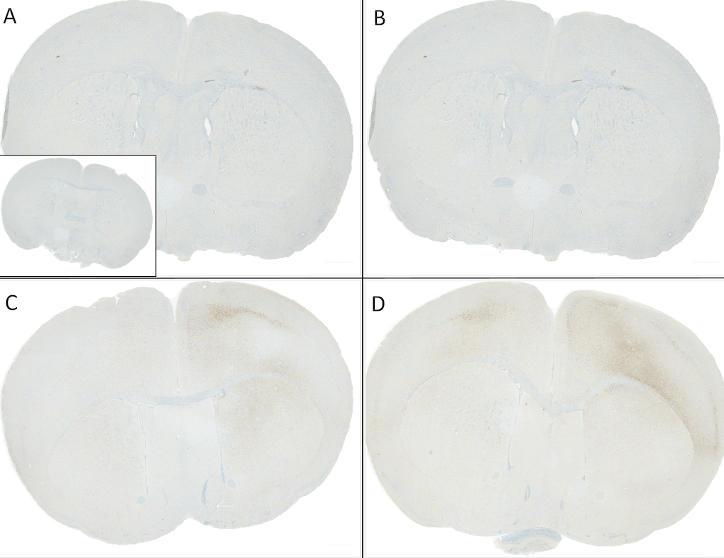
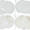
<p>C57/BL6 mice were injected with sonicated recombinant mouse alpha synuclein monomers or fibrils at 8 weeks of age. Mice were unilaterally injected in the dorsal striatum (bregma AP + 0.2 mm, L +/1 2.0 mm, V – 3.0 mm) and sacrificed 30 days post-injection.  (A) 1.25 uL  mouse alpha synuclein monomers (SPR-323). (B) 2.5 uL mouse alpha synuclein monomers (SPR-323). (C) 2.5 ug alpha synuclein PFFs (SPR-324). (D) 5 ug alpha synuclein PFFs (SPR-324)  Inset: PBS (negative control). Primary antibody: Anti-Alpha Synuclein pSer129 (SMC-600) at 1:10 000. Secondary antibody: anti-rabbit HRP. Mice injected with PFF displayed alpha synuclein staining in the striatum and cortex and contralateral to the injection site. Courtesy of: Porsolt.</p>

C57/BL6 mice were injected with sonicated recombinant mouse alpha synuclein monomers or fibrils at 8 weeks of age. Mice were unilaterally injected in the dorsal striatum (bregma AP + 0.2 mm, L +/1 2.0 mm, V – 3.0 mm) and sacrificed 30 days post-injection. (A) 1.25 uL mouse alpha synuclein monomers (SPR-323). (B) 2.5 uL mouse alpha synuclein monomers (SPR-323). (C) 2.5 ug alpha synuclein PFFs (SPR-324). (D) 5 ug alpha synuclein PFFs (SPR-324) Inset: PBS (negative control). Primary antibody: Anti-Alpha Synuclein pSer129 (SMC-600) at 1:10 000. Secondary antibody: anti-rabbit HRP. Mice injected with PFF displayed alpha synuclein staining in the striatum and cortex and contralateral to the injection site. Courtesy of: Porsolt.
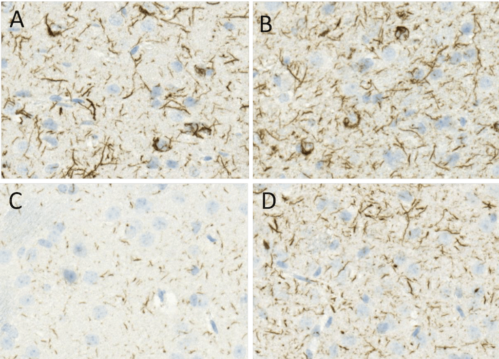
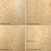
<p>C57/BL6 mice were injected with 5 ug sonicated mouse recombinant alpha synuclein PFFs (SPR-324) at 8 weeks of age. Mice were unilaterally injected in the dorsal striatum (bregma AP + 0.2 mm, L +/1 2.0 mm, V – 3.0 mm) and sacrificed 30 days post-injection. (A) contralateral cortex. (B) ipsilateral cortex. (C) contralateral striatum. (D) ipsilateral striatum. Primary antibody: Anti-Alpha Synuclein pSer129 (SMC-600) at 1:10 000. Secondary antibody: anti-rabbit HRP. Mice injected with PFF displayed alpha synuclein staining in the striatum and cortex and contralateral to the injection site. Courtesy of: Porsolt.</p>

C57/BL6 mice were injected with 5 ug sonicated mouse recombinant alpha synuclein PFFs (SPR-324) at 8 weeks of age. Mice were unilaterally injected in the dorsal striatum (bregma AP + 0.2 mm, L +/1 2.0 mm, V – 3.0 mm) and sacrificed 30 days post-injection. (A) contralateral cortex. (B) ipsilateral cortex. (C) contralateral striatum. (D) ipsilateral striatum. Primary antibody: Anti-Alpha Synuclein pSer129 (SMC-600) at 1:10 000. Secondary antibody: anti-rabbit HRP. Mice injected with PFF displayed alpha synuclein staining in the striatum and cortex and contralateral to the injection site. Courtesy of: Porsolt.

Reviews

1 review for Alpha Synuclein Protein

  1. 5 out of 5

    :

    Based on validation through cited publications.

Add a review

Your email address will not be published. Required fields are marked *